
Search Count: 48
 |
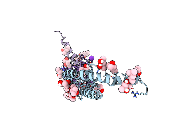
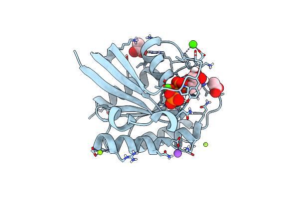
Structure Of A K+ Selective Nak Mutant (Nak2K, Laue Diffraction) In The Presence Of An Electric Field Of ~0.8Mv/Cm Along The Crystallographic Z Axis, 500Ns, With Eightfold Extrapolation Of Structure Factor Differences
Organism: Bacillus cereus m1550
Method: X-RAY DIFFRACTION Resolution:2.01 Å Release Date: 2023-07-26 Classification: MEMBRANE PROTEIN Ligands: MPD, K |
 |
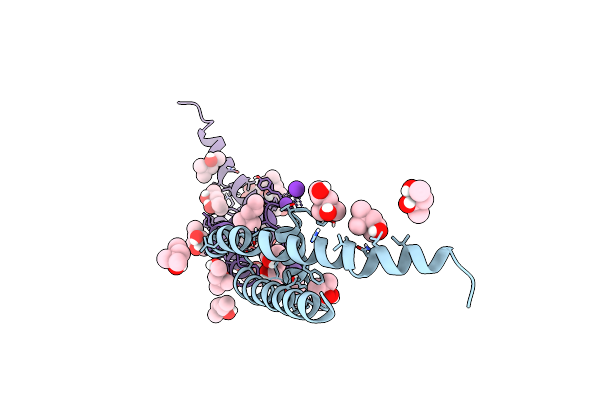
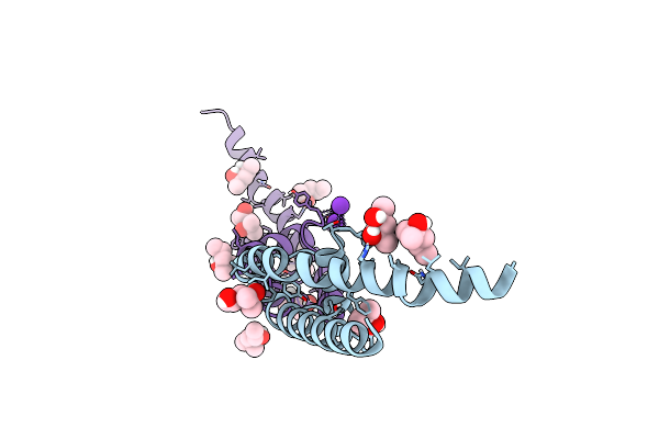
Structure Of A K+ Selective Nak Mutant (Nak2K, Laue Diffraction) In The Presence Of An Electric Field Of ~0.8Mv/Cm Along The Crystallographic Z Axis, 100Ns, With Eightfold Extrapolation Of Structure Factor Differences
Organism: Bacillus cereus m1550
Method: X-RAY DIFFRACTION Resolution:2.01 Å Release Date: 2023-07-26 Classification: MEMBRANE PROTEIN Ligands: MPD, K |
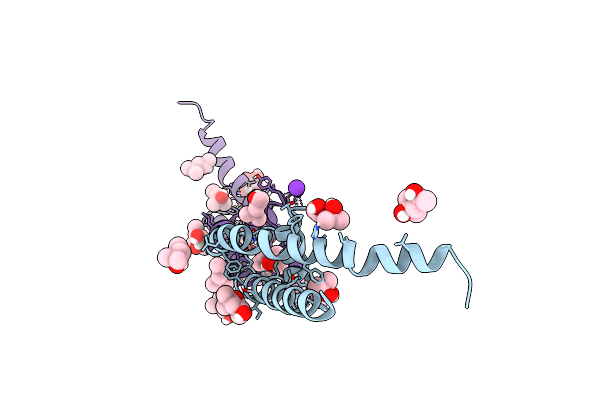 |
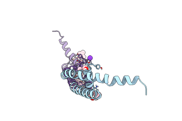
Structure Of A K+ Selective Nak Mutant (Nak2K, Laue Diffraction) In The Presence Of An Electric Field Of ~0.8Mv/Cm Along The Crystallographic Z Axis, 200Ns, With Eightfold Extrapolation Of Structure Factor Differences
Organism: Bacillus cereus m1550
Method: X-RAY DIFFRACTION Resolution:2.01 Å Release Date: 2023-07-26 Classification: MEMBRANE PROTEIN Ligands: MPD, K |
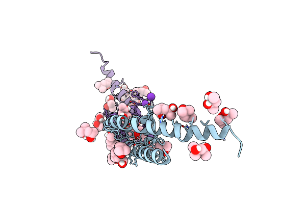 |
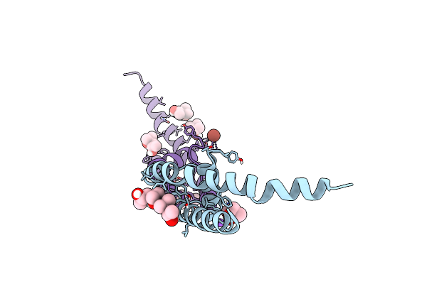
Structure Of A K+ Selective Nak Mutant (Nak2K, Laue Diffraction) In The Presence Of An Electric Field Of ~0.8Mv/Cm Along The Crystallographic Z Axis, 1Us, With Eightfold Extrapolation Of Structure Factor Differences
Organism: Bacillus cereus
Method: X-RAY DIFFRACTION Resolution:2.01 Å Release Date: 2023-07-26 Classification: MEMBRANE PROTEIN Ligands: MPD, K |
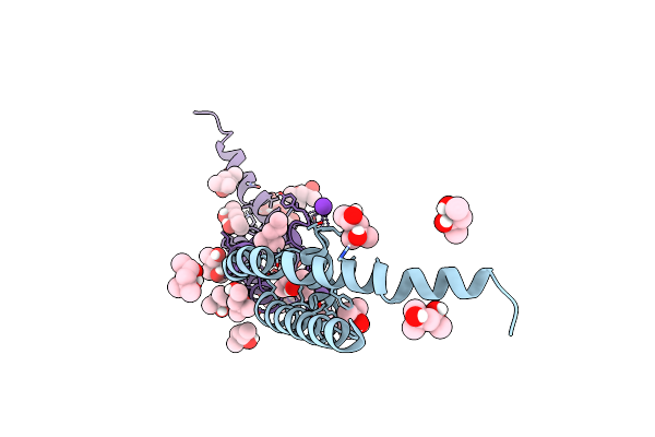 |
Structure Of A K+ Selective Nak Mutant (Nak2K, Laue Diffraction, No Electric Field)
Organism: Bacillus cereus m1550
Method: X-RAY DIFFRACTION Resolution:2.01 Å Release Date: 2023-06-14 Classification: MEMBRANE PROTEIN Ligands: MPD, K |
 |
Organism: Bacillus cereus m1550
Method: X-RAY DIFFRACTION Resolution:1.60 Å Release Date: 2023-06-14 Classification: MEMBRANE PROTEIN Ligands: MPD, K |
 |
Organism: Bacillus cereus m1550
Method: X-RAY DIFFRACTION Resolution:2.05 Å Release Date: 2023-06-14 Classification: MEMBRANE PROTEIN Ligands: MPD, K |
 |
Organism: Bacillus cereus m1550
Method: X-RAY DIFFRACTION Resolution:1.65 Å Release Date: 2023-06-14 Classification: MEMBRANE PROTEIN Ligands: MPD, K, GAL |
 |
Organism: Bacillus cereus m1550
Method: X-RAY DIFFRACTION Resolution:2.10 Å Release Date: 2023-06-14 Classification: MEMBRANE PROTEIN Ligands: MPD, TL, NA |
 |
Organism: Bacillus cereus m1550
Method: X-RAY DIFFRACTION Resolution:2.00 Å Release Date: 2023-06-14 Classification: MEMBRANE PROTEIN Ligands: MPD, TL, NA |
 |
Organism: Bacillus cereus m1550
Method: X-RAY DIFFRACTION Resolution:1.99 Å Release Date: 2023-06-14 Classification: MEMBRANE PROTEIN Ligands: TL, K |
 |
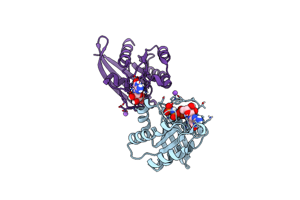
Organism: Homo sapiens
Method: X-RAY DIFFRACTION Resolution:1.65 Å Release Date: 2017-07-19 Classification: ONCOPROTEIN, HYDROLASE Ligands: CA, MG, NA, GNP |
 |
Organism: Homo sapiens
Method: X-RAY DIFFRACTION Resolution:1.35 Å Release Date: 2017-07-19 Classification: ONCOPROTEIN, HYDROLASE Ligands: CA, GNP, MG |
 |
Organism: Homo sapiens
Method: X-RAY DIFFRACTION Resolution:1.25 Å Release Date: 2017-07-19 Classification: ONCOPROTEIN, HYDROLASE Ligands: GNP, CA, MG, NA, ACT |
 |
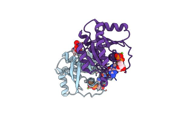
Organism: Salpingoeca rosetta
Method: X-RAY DIFFRACTION Resolution:1.60 Å Release Date: 2017-07-19 Classification: ONCOPROTEIN, HYDROLASE Ligands: MG, NA, GNP |
 |
Organism: Salpingoeca rosetta
Method: X-RAY DIFFRACTION Resolution:1.85 Å Release Date: 2017-07-19 Classification: ONCOPROTEIN, HYDROLASE Ligands: GDP, MG, GOL |
 |
Organism: Rattus norvegicus
Method: X-RAY DIFFRACTION Resolution:1.80 Å Release Date: 2017-01-11 Classification: PEPTIDE BINDING PROTEIN Ligands: GOL |
 |
Organism: Rattus norvegicus
Method: X-RAY DIFFRACTION Resolution:1.75 Å Release Date: 2017-01-11 Classification: PEPTIDE BINDING PROTEIN Ligands: GOL |
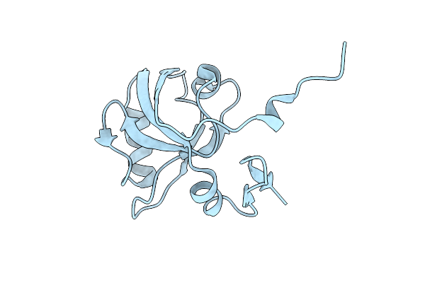 |
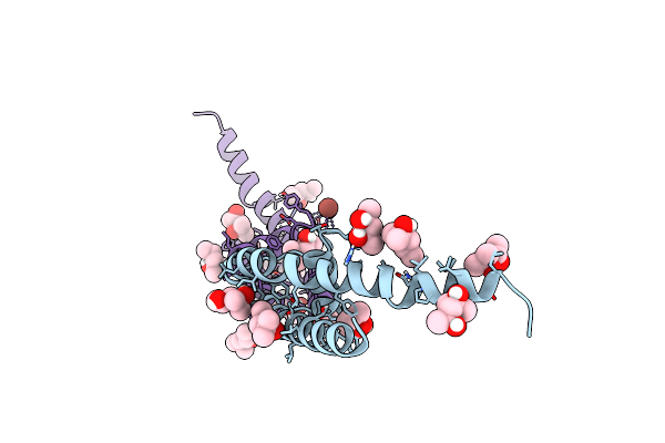
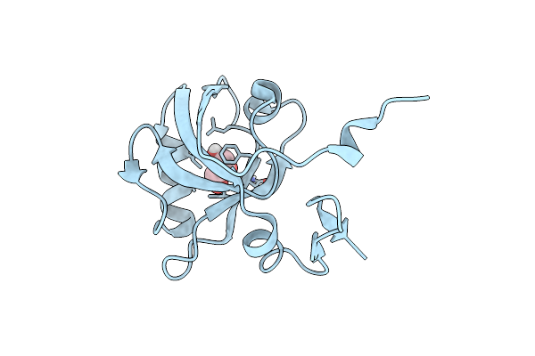
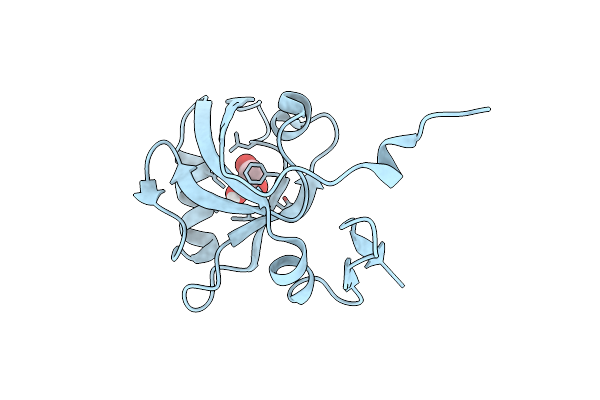
The Third Pdz Domain From The Synaptic Protein Psd-95 (G330T, H372A Double Mutant)
Organism: Rattus norvegicus
Method: X-RAY DIFFRACTION Resolution:1.60 Å Release Date: 2017-01-11 Classification: PEPTIDE BINDING PROTEIN |
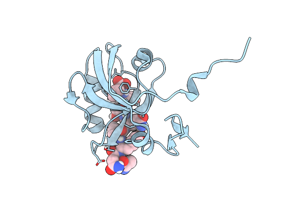 |
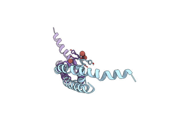
The Third Pdz Domain From The Synaptic Protein Psd-95 (G330T, H372A Double Mutant) In Complex With A C-Terminal Peptide Derived From Cript
Organism: Rattus norvegicus
Method: X-RAY DIFFRACTION Resolution:1.80 Å Release Date: 2017-01-11 Classification: PEPTIDE BINDING PROTEIN |