
Search Count: 313
 |
Organism: Serratia marcescens
Method: X-RAY DIFFRACTION Release Date: 2025-10-22 Classification: SIGNALING PROTEIN |
 |
Organism: Serratia marcescens
Method: X-RAY DIFFRACTION Release Date: 2025-10-22 Classification: SIGNALING PROTEIN |
 |
Organism: Serratia marcescens
Method: X-RAY DIFFRACTION Release Date: 2025-10-22 Classification: SIGNALING PROTEIN |
 |
Crystal Structure Of Gdp-Bound Kras G12D/M67R: Suppressing G12D Oncogenicity Via Second-Site M67R Mutation
Organism: Homo sapiens
Method: X-RAY DIFFRACTION Release Date: 2025-10-08 Classification: ONCOPROTEIN Ligands: GDP, MG |
 |
Crystal Structure Of Gdp-Bound Kras G12D/I55E: Suppressing G12D Oncogenicity Via Second-Site I55E Mutation
Organism: Homo sapiens
Method: X-RAY DIFFRACTION Release Date: 2025-10-08 Classification: ONCOPROTEIN Ligands: GDP, MG |
 |
Crystal Structure Of Gdp-Bound Kras G12D/F28K: Suppressing G12D Oncogenicity Via Second-Site F28K Mutation
Organism: Homo sapiens
Method: X-RAY DIFFRACTION Release Date: 2025-10-08 Classification: ONCOPROTEIN Ligands: GDP, MG, SO4 |
 |
Crystal Structure Of Gdp-Bound Kras G12D/P34R: Suppressing G12D Oncogenicity Via Second-Site P34R Mutation
Organism: Homo sapiens
Method: X-RAY DIFFRACTION Release Date: 2025-10-08 Classification: ONCOPROTEIN Ligands: MG, GDP |
 |
Crystal Structure Of Gdp-Bound Kras G12D/R41Q: Suppressing G12D Oncogenicity Via Second-Site R41Q Mutation
Organism: Homo sapiens
Method: X-RAY DIFFRACTION Release Date: 2025-10-08 Classification: ONCOPROTEIN Ligands: GDP, MG |
 |
Crystal Structure Of Gdp-Bound Kras G12D/V45E: Suppressing G12D Oncogenicity Via Second-Site V45E Mutation
Organism: Homo sapiens
Method: X-RAY DIFFRACTION Release Date: 2025-10-08 Classification: ONCOPROTEIN Ligands: GDP, MG |
 |
Crystal Structure Of Gdp-Bound Kras G12D/D54R: Suppressing G12D Oncogenicity Via Second-Site D54R Mutation
Organism: Homo sapiens
Method: X-RAY DIFFRACTION Release Date: 2025-10-08 Classification: ONCOPROTEIN Ligands: GDP, MG, SO4 |
 |
Crystal Structure Of Gdp-Bound Kras G12D/G60R: Suppressing G12D Oncogenicity Via Second-Site G60R Mutation
Organism: Homo sapiens
Method: X-RAY DIFFRACTION Release Date: 2025-10-08 Classification: ONCOPROTEIN Ligands: MG, GDP |
 |
Crystal Structure Of Gdp-Bound Kras G12D/V103Y: Suppressing G12D Oncogenicity Via Second-Site V103Y Mutation
Organism: Homo sapiens
Method: X-RAY DIFFRACTION Release Date: 2025-10-08 Classification: ONCOPROTEIN Ligands: MG, GDP |
 |
Crystal Structure Of Gdp-Bound Kras G12D/E62Q: Suppressing G12D Oncogenicity Via Second-Site E62Q Mutation
Organism: Homo sapiens
Method: X-RAY DIFFRACTION Release Date: 2025-10-08 Classification: ONCOPROTEIN Ligands: MG, GDP, SO4 |
 |
Crystal Structure Of Gdp-Bound Kras E3K/G12D: Suppressing G12D Oncogenicity Via Second-Site E3K Mutation
Organism: Homo sapiens
Method: X-RAY DIFFRACTION Release Date: 2025-10-08 Classification: ONCOPROTEIN Ligands: MG, GDP, GOL |
 |
Organism: Homo sapiens
Method: X-RAY DIFFRACTION Release Date: 2025-07-30 Classification: HYDROLASE Ligands: CL, ZN, MES, EDO, PEG, MPD |
 |
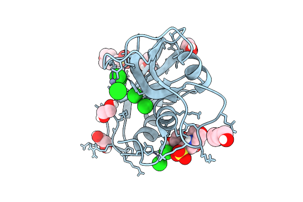
Crystal Structure Of Human Ribonuclease 7 (Rnase 7) In Complex With 5'-Adenosine Monophosphate (5'-Amp)
Organism: Homo sapiens
Method: X-RAY DIFFRACTION Release Date: 2025-07-30 Classification: HYDROLASE Ligands: BO3, AMP, GOL, EDO |
 |
Organism: Gibberella zeae
Method: X-RAY DIFFRACTION Resolution:1.58 Å Release Date: 2025-04-23 Classification: TRANSCRIPTION |
 |
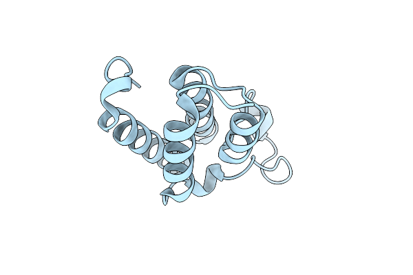
Organism: Homo sapiens
Method: ELECTRON MICROSCOPY Release Date: 2025-03-05 Classification: PROTEIN BINDING |
 |
Crystal Structure Of Xanthomonas Oryzae Pv. Oryzae Nadh-Quinone Oxidoreductase Subunits Nuoe And Nuof
Organism: Xanthomonas oryzae pv. oryzae
Method: X-RAY DIFFRACTION Release Date: 2024-12-04 Classification: OXIDOREDUCTASE Ligands: GOL, FMN, FES |
 |
Crystal Structure Of Sars Cov-2 Papain-Like Protease Plpro-C111S In Complex With Gznl-P4
Organism: Severe acute respiratory syndrome coronavirus 2
Method: X-RAY DIFFRACTION Resolution:2.31 Å Release Date: 2024-12-04 Classification: HYDROLASE Ligands: ZN, A1LZ5 |