
Search Count: 44
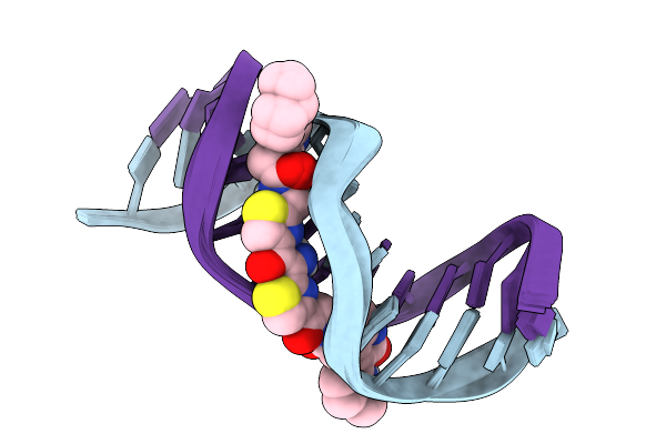 |
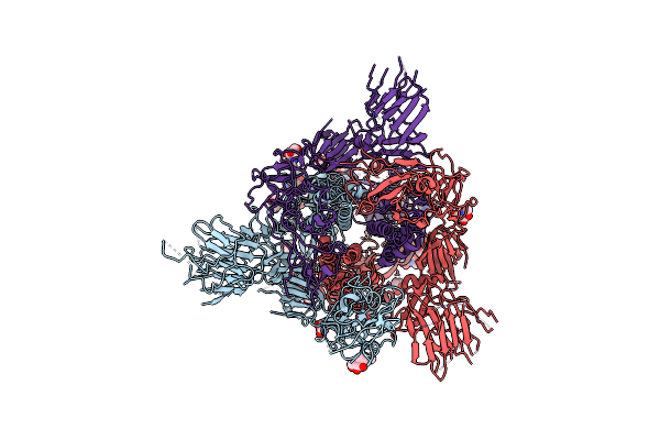
The Specificity And Structure Of Dna Crosslinking By A Gut Bacterial Genotoxin
Organism: Escherichia coli
Method: SOLUTION NMR Release Date: 2025-11-19 Classification: DNA Ligands: A1CEC |
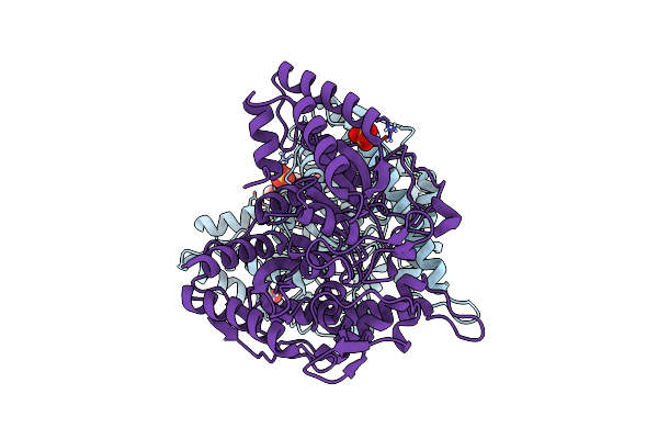 |
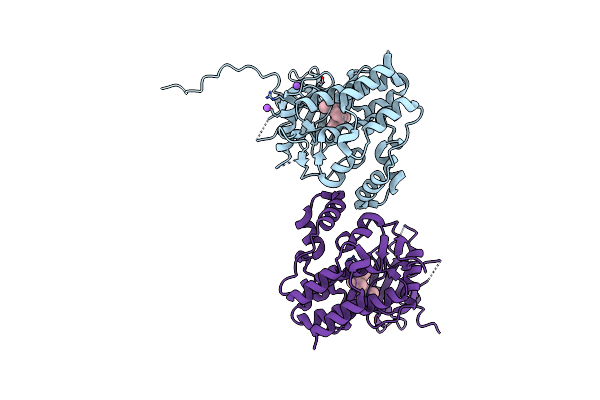
Organism: Uncultured bacterium
Method: X-RAY DIFFRACTION Resolution:2.32 Å Release Date: 2025-02-05 Classification: HYDROLASE Ligands: PG6, SO4 |
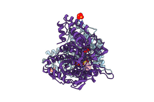 |
Organism: Uncultured bacterium
Method: X-RAY DIFFRACTION Resolution:2.18 Å Release Date: 2025-02-05 Classification: HYDROLASE Ligands: SO4, PMS |
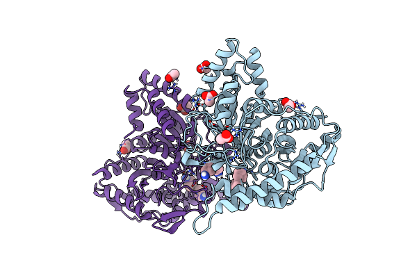 |
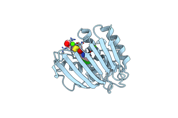
Organism: Uncultured bacterium
Method: X-RAY DIFFRACTION Resolution:2.00 Å Release Date: 2025-02-05 Classification: HYDROLASE Ligands: ACT, A1IHK |
 |
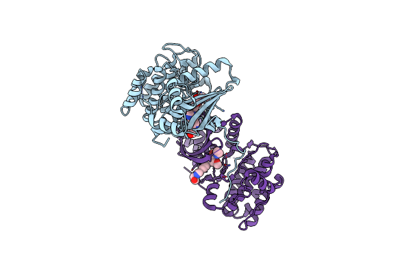
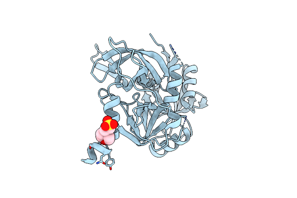
Crystal Structure Of A Novel Pu Plastic Degradation Urethanase Umg-Sp2 Mutant From Uncultured Bacterium In Complex With Ligand
Organism: Metagenome
Method: X-RAY DIFFRACTION Resolution:2.40 Å Release Date: 2025-01-15 Classification: HYDROLASE Ligands: A1D5H, GOL |
 |
Organism: Severe acute respiratory syndrome coronavirus 2
Method: ELECTRON MICROSCOPY Release Date: 2024-12-04 Classification: VIRAL PROTEIN Ligands: NAG |
 |
Organism: Homo sapiens
Method: X-RAY DIFFRACTION Resolution:1.75 Å Release Date: 2024-11-06 Classification: SIGNALING PROTEIN Ligands: A1H5R, NA |
 |
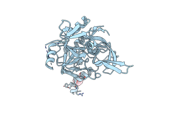
Crystal Structure Of A Novel Pu Plastic Degradation Urethanase Umg-Sp2 From Uncultured Bacterium
Organism: Uncultured bacterium
Method: X-RAY DIFFRACTION Resolution:2.16 Å Release Date: 2024-09-18 Classification: HYDROLASE Ligands: GOL, SO4, PMS |
 |
Organism: Homo sapiens
Method: X-RAY DIFFRACTION Resolution:3.10 Å Release Date: 2024-01-10 Classification: SIGNALING PROTEIN Ligands: VRU |
 |
Pseudomonas Aeruginosa Dna Gyrase B 24Kda Atpase Subdomain Complexed With Ebl3021
Organism: Pseudomonas aeruginosa pao1
Method: X-RAY DIFFRACTION Resolution:1.60 Å Release Date: 2023-03-29 Classification: DNA BINDING PROTEIN Ligands: CA, R53 |
 |
Pseudomonas Aeruginosa Dna Gyrase B 24Kda Atpase Subdomain Complexed With Novobiocin
Organism: Pseudomonas aeruginosa (strain atcc 15692 / dsm 22644 / cip 104116 / jcm 14847 / lmg 12228 / 1c / prs 101 / pao1)
Method: X-RAY DIFFRACTION Resolution:1.32 Å Release Date: 2022-10-05 Classification: DNA BINDING PROTEIN Ligands: NOV, MRD, MES |
 |
Pseudomonas Aeruginosa Dna Gyrase B 24Kda Atpase Subdomain Complexed With Ebl2888
Organism: Pseudomonas aeruginosa (strain atcc 15692 / dsm 22644 / cip 104116 / jcm 14847 / lmg 12228 / 1c / prs 101 / pao1)
Method: X-RAY DIFFRACTION Resolution:2.20 Å Release Date: 2022-10-05 Classification: DNA BINDING PROTEIN Ligands: 80S |
 |
Acinetobacter Baumannii Dna Gyrase B 23Kda Atpase Subdomain Complexed With Novobiocin
Organism: Acinetobacter baumannii 1419130
Method: X-RAY DIFFRACTION Resolution:1.90 Å Release Date: 2022-09-28 Classification: DNA BINDING PROTEIN Ligands: NOV, EDO |
 |
Acinetobacter Baumannii Dna Gyrase B 23Kda Atpase Subdomain Complexed With Ebl2704
Organism: Acinetobacter baumannii 1419130
Method: X-RAY DIFFRACTION Resolution:1.60 Å Release Date: 2022-09-28 Classification: DNA BINDING PROTEIN Ligands: 80S, 81N |
 |
Acinetobacter Baumannii Dna Gyrase B 23Kda Atpase Subdomain Complexed With Ebl2888
Organism: Acinetobacter baumannii 1419130
Method: X-RAY DIFFRACTION Resolution:1.55 Å Release Date: 2022-09-28 Classification: DNA BINDING PROTEIN Ligands: 80S, CA |
 |
Organism: Escherichia coli (strain k12)
Method: X-RAY DIFFRACTION Resolution:1.16 Å Release Date: 2022-07-20 Classification: DNA BINDING PROTEIN Ligands: PO4, N1N |
 |
Organism: Escherichia coli (strain k12)
Method: X-RAY DIFFRACTION Resolution:1.65 Å Release Date: 2022-07-20 Classification: DNA BINDING PROTEIN Ligands: 4QR, TRS, MG |
 |
Crystal Structure Of Peruvianin-I (Cysteine Peptidase From Thevetia Peruviana Latex)
Organism: Thevetia peruviana
Method: X-RAY DIFFRACTION Resolution:2.15 Å Release Date: 2020-05-06 Classification: HYDROLASE |
 |
Organism: Homo sapiens
Method: X-RAY DIFFRACTION Resolution:1.65 Å Release Date: 2017-10-25 Classification: HYDROLASE Ligands: MES |
 |
Organism: Homo sapiens
Method: X-RAY DIFFRACTION Resolution:1.70 Å Release Date: 2017-10-25 Classification: HYDROLASE Ligands: MES |