
Search Count: 6
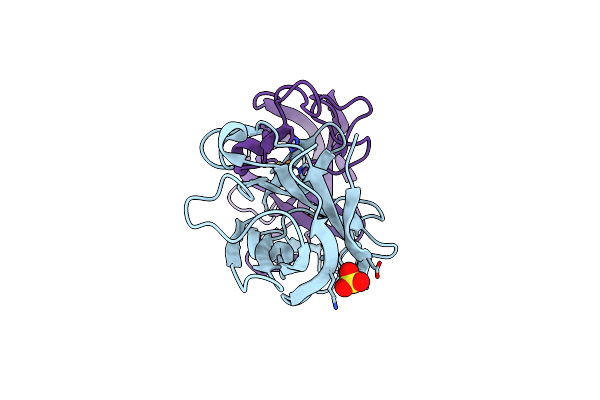 |
Crystal Structure Analysis Of The As-Solated P19 Protein From Campylobacter Jejuni At 1.45 A At Ph 9.0
Organism: Campylobacter jejuni
Method: X-RAY DIFFRACTION Resolution:1.45 Å Release Date: 2010-07-21 Classification: TRANSPORT PROTEIN Ligands: CU, SO4 |
 |
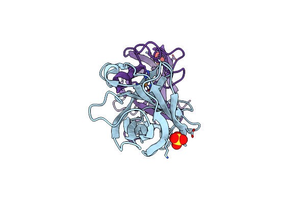
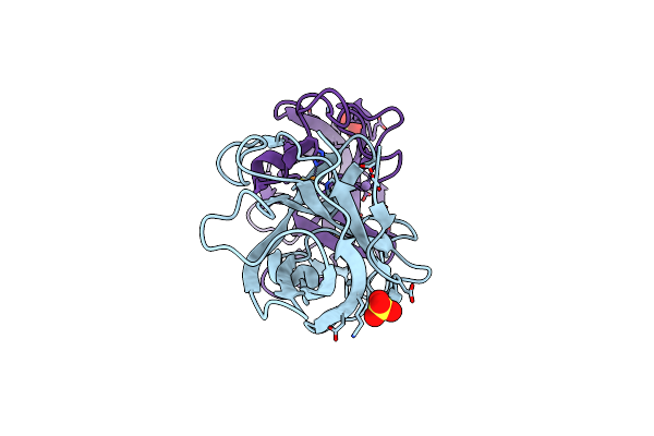
Crystal Structure Analysis Of The Apo P19 Protein From Campylobacter Jejuni At 1.59 A At Ph 9
Organism: Campylobacter jejuni
Method: X-RAY DIFFRACTION Resolution:1.59 Å Release Date: 2010-07-21 Classification: TRANSPORT PROTEIN Ligands: ZN, SO4 |
 |
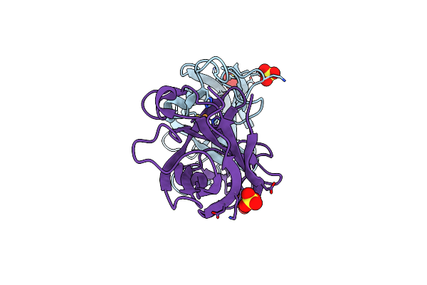
Crystal Structure Analysis Of The Copper-Reconstituted P19 Protein From Campylobacter Jejuni At 1.65 A At Ph 10.0
Organism: Campylobacter jejuni
Method: X-RAY DIFFRACTION Resolution:1.65 Å Release Date: 2010-07-21 Classification: TRANSPORT PROTEIN Ligands: CU, SO4 |
 |
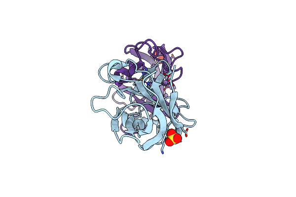
Crystal Structure Analysis Of The 'As-Isolated' P19 Protein From Campylobacter Jejuni At 1.65 A At Ph 9.0
Organism: Campylobacter jejuni
Method: X-RAY DIFFRACTION Resolution:1.65 Å Release Date: 2010-07-21 Classification: TRANSPORT PROTEIN Ligands: CU, SO4 |
 |
Crystal Structure Analysis Of Manganese Treated P19 Protein From Campylobacter Jejuni At 1.41 A At Ph 9
Organism: Campylobacter jejuni
Method: X-RAY DIFFRACTION Resolution:1.41 Å Release Date: 2010-07-21 Classification: TRANSPORT PROTEIN Ligands: CU, MN, SO4 |
 |
Crystal Structure Analysis Of Manganese Treated P19 Protein From Campylobacter Jejuni At 2.73 A At Ph 9 And Manganese Peak Wavelength (1.893 A)
Organism: Campylobacter jejuni
Method: X-RAY DIFFRACTION Resolution:2.73 Å Release Date: 2010-07-21 Classification: TRANSPORT PROTEIN Ligands: CU, MN, SO4 |