
Search Count: 37
 |
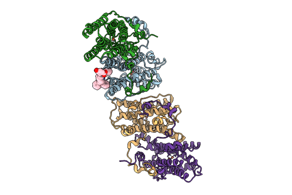
Organism: Trypanosoma brucei brucei
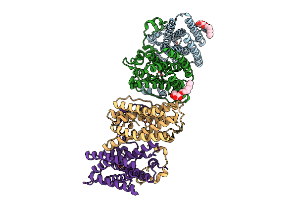
Method: X-RAY DIFFRACTION Release Date: 2025-09-10 Classification: OXIDOREDUCTASE Ligands: FE, OH, BOG |
 |
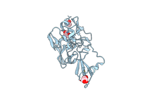
The Crystal Structure Of The Trypanosome Alternative Oxidase Complexed With A Trypanocidal Phosphonium Derivative (Compound1)
Organism: Trypanosoma brucei brucei
Method: X-RAY DIFFRACTION Release Date: 2025-09-10 Classification: OXIDOREDUCTASE Ligands: FE, OH, A1L8E |
 |
Organism: Aplysia kurodai
Method: X-RAY DIFFRACTION Resolution:2.70 Å Release Date: 2023-11-15 Classification: HYDROLASE Ligands: NAG |
 |
Organism: Aplysia kurodai
Method: X-RAY DIFFRACTION Resolution:1.15 Å Release Date: 2023-11-15 Classification: UNKNOWN FUNCTION Ligands: GOL |
 |
Organism: Aplysia kurodai
Method: X-RAY DIFFRACTION Resolution:1.40 Å Release Date: 2023-11-15 Classification: UNKNOWN FUNCTION Ligands: GOL, ACE |
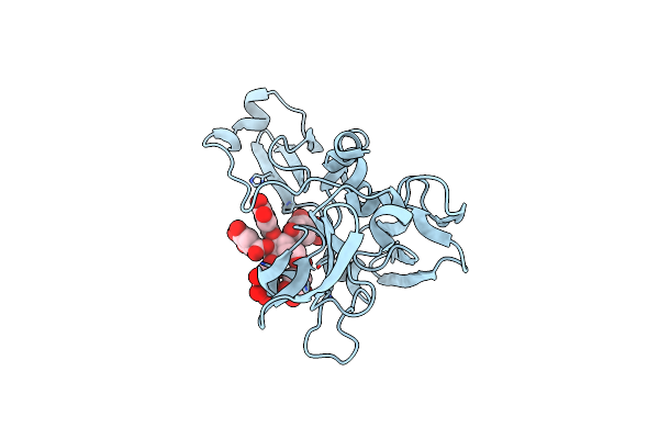 |
Eisenia Hydrolysis-Enhancing Protein From Aplysia Kurodai Complex With Tannic Acid
Organism: Aplysia kurodai
Method: X-RAY DIFFRACTION Resolution:1.90 Å Release Date: 2023-11-15 Classification: UNKNOWN FUNCTION Ligands: GGP |
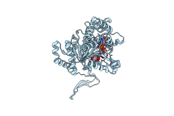 |
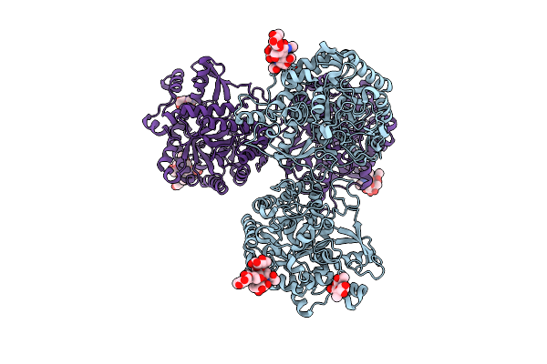
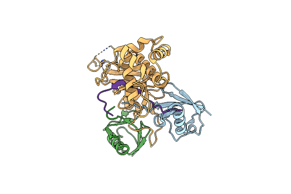
Crystal Structure Of Isocitrate Dehydrogenase D252N Mutant From Trypanosoma Brucei In Complexed With Nadp+, Alpha-Ketoglutarate, And Ca2+
Organism: Trypanosoma brucei brucei (strain 927/4 gutat10.1)
Method: X-RAY DIFFRACTION Resolution:1.90 Å Release Date: 2022-02-16 Classification: OXIDOREDUCTASE Ligands: NAP, CA, ICT |
 |
 |
 |
 |
 |
 |
 |
 |
 |
 |
Organism: Aequorea victoria
Method: X-RAY DIFFRACTION Resolution:3.20 Å Release Date: 2017-12-13 Classification: FLUORESCENT PROTEIN |
 |
Crystal Structure Of Dnmt1 Rfts Domain In Complex With K18/K23 Mono-Ubiquitylated Histone H3
Organism: Homo sapiens
Method: X-RAY DIFFRACTION Resolution:2.00 Å Release Date: 2017-11-15 Classification: SIGNALING PROTEIN/TRANSFERASE Ligands: ZN |
 |
Organism: Zymomonas mobilis
Method: X-RAY DIFFRACTION Resolution:1.67 Å Release Date: 2017-10-18 Classification: CELL ADHESION Ligands: TPP, MG, SO4, EDO |
 |
Organism: Saccharomyces cerevisiae (strain awri796), Saccharomyces cerevisiae (strain atcc 204508 / s288c), Homo sapiens, Saccharomyces cerevisiae (strain fostersb)
Method: X-RAY DIFFRACTION Resolution:2.20 Å Release Date: 2017-08-16 Classification: GTP BINDING PROTEIN Ligands: GTP, MG |