
Search Count: 19
 |
Organism: Homo sapiens
Method: X-RAY DIFFRACTION Resolution:1.44 Å Release Date: 2025-01-22 Classification: IMMUNE SYSTEM Ligands: TB, 9JE, BTB, PRO, YT3, PG4, ER3 |
 |
Organism: Neisseria gonorrhoeae f62
Method: X-RAY DIFFRACTION Resolution:2.45 Å Release Date: 2024-09-11 Classification: ELECTRON TRANSPORT Ligands: HEC, SO4 |
 |
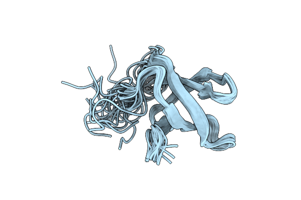
Organism: Homo sapiens
Method: SOLUTION NMR Release Date: 2020-12-23 Classification: LIPID BINDING PROTEIN Ligands: ZN |
 |
Organism: Saccharomyces cerevisiae
Method: X-RAY DIFFRACTION Resolution:2.03 Å Release Date: 2020-09-16 Classification: STRUCTURAL PROTEIN Ligands: SO4 |
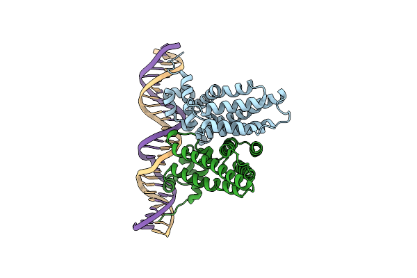 |
Tetr Family Transcriptional Regulator Cifr C99T-C107S-C181R Cysteines Mutant Complexed With 26Bp Double-Strand Operator Dna
Organism: Pseudomonas aeruginosa ucbpp-pa14, Pseudomonas aeruginosa
Method: X-RAY DIFFRACTION Resolution:2.80 Å Release Date: 2020-01-29 Classification: TRANSCRIPTION/DNA |
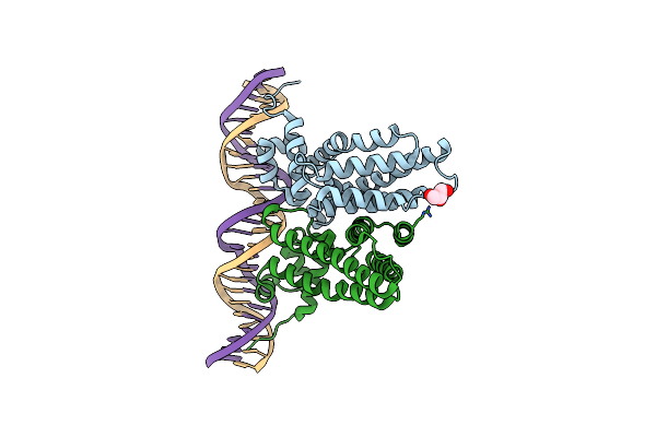 |
Tetr Family Transcriptional Regulator Cifr C99T-C181R Cysteines Mutant Complexed With 26Bp Double-Strand Operator Dna
Organism: Pseudomonas aeruginosa
Method: X-RAY DIFFRACTION Resolution:2.60 Å Release Date: 2020-01-29 Classification: TRANSCRIPTION/DNA Ligands: GOL |
 |
Tetr Family Transcriptional Regulator Cifr C99T-C181R Cysteine Mutant Complexed With 26Bp Double-Strand Operator Dna And Apo-Cifr C99T-C181R
Organism: Pseudomonas aeruginosa
Method: X-RAY DIFFRACTION Resolution:3.00 Å Release Date: 2020-01-29 Classification: TRANSCRIPTION/DNA |
 |
Organism: Pseudomonas aeruginosa
Method: X-RAY DIFFRACTION Resolution:1.85 Å Release Date: 2019-11-20 Classification: ELECTRON TRANSPORT Ligands: HEC |
 |
Organism: Prochlorococcus marinus (strain mit 9303)
Method: SOLUTION NMR Release Date: 2019-08-21 Classification: RNA BINDING PROTEIN |
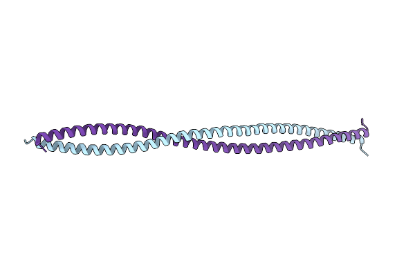 |
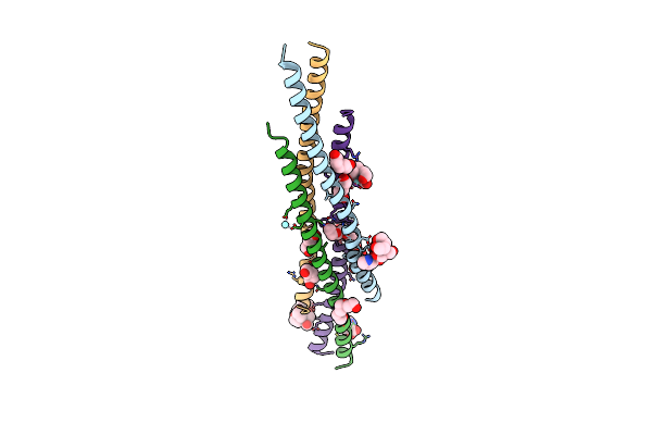
Structure Of The I65M Mutant Of Nemo(51-112) With N- And C-Terminal Coiled-Coil Adaptors.
Organism: Homo sapiens
Method: X-RAY DIFFRACTION Resolution:2.50 Å Release Date: 2019-08-07 Classification: TRANSCRIPTION |
 |
Organism: Homo sapiens
Method: X-RAY DIFFRACTION Resolution:1.78 Å Release Date: 2019-07-31 Classification: TRANSCRIPTION |
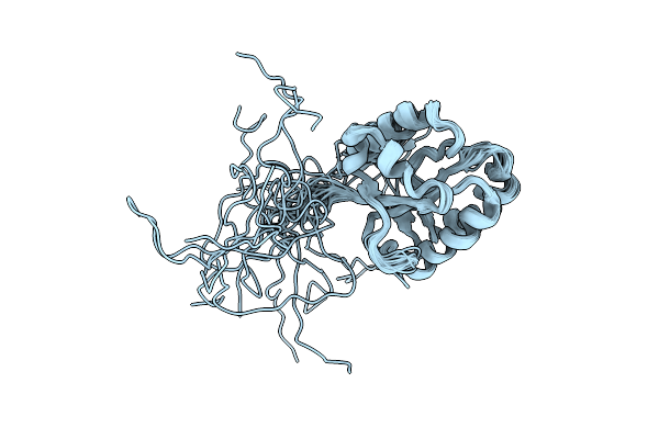 |
Organism: Saccharomyces cerevisiae
Method: SOLUTION NMR Release Date: 2018-07-04 Classification: PROTEIN TRANSPORT |
 |
Organism: Lachancea thermotolerans
Method: X-RAY DIFFRACTION Resolution:2.29 Å Release Date: 2013-03-20 Classification: UNKNOWN FUNCTION Ligands: SO4 |
 |
Organism: Lachancea thermotolerans cbs 6340
Method: X-RAY DIFFRACTION Resolution:3.06 Å Release Date: 2012-12-26 Classification: PROTEIN TRANSPORT |
 |
Organism: Kluyveromyces lactis
Method: X-RAY DIFFRACTION Resolution:3.00 Å Release Date: 2012-07-04 Classification: TRANSPORT PROTEIN Ligands: SO4 |
 |
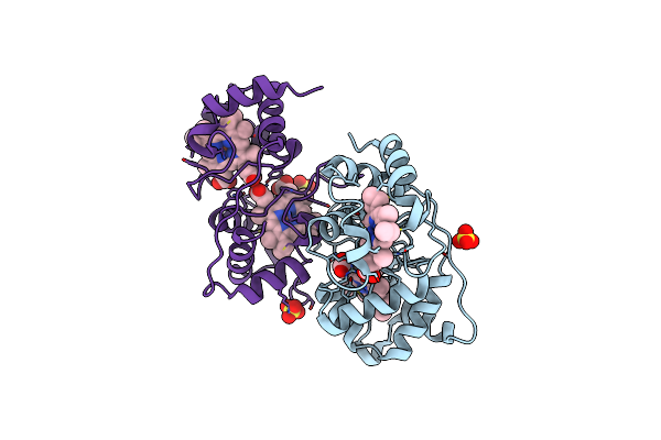
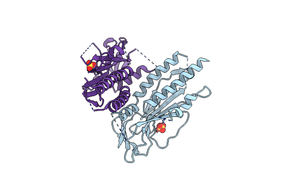
Crystal Structure Of A Complex Between Protein Phosphatase 1 Alpha (Pp1) And The Pp1 Binding And Pdz Domains Of Spinophilin
Organism: Homo sapiens, Rattus norvegicus
Method: X-RAY DIFFRACTION Resolution:1.85 Å Release Date: 2010-03-23 Classification: HYDROLASE Ligands: GOL, MN, MES |
 |
Crystal Structure Of A Complex Between Protein Phosphatase 1 Alpha (Pp1), The Pp1 Binding And Pdz Domains Of Spinophilin And The Small Natural Molecular Toxin Nodularin-R
Organism: Homo sapiens, Rattus norvegicus, Nodularia spumigena
Method: X-RAY DIFFRACTION Resolution:2.00 Å Release Date: 2010-03-23 Classification: HYDROLASE/HYDROLASE INHIBITOR Ligands: GOL, MN |
 |
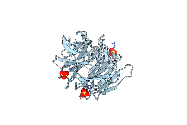
Crystal Structure Of A Complex Between Protein Phosphatase 1 Alpha (Pp1) And The Pp1 Binding And Pdz Domains Of Neurabin
Organism: Homo sapiens, Rattus norvegicus
Method: X-RAY DIFFRACTION Resolution:2.20 Å Release Date: 2010-03-23 Classification: HYDROLASE/HYDROLASE REGULATOR Ligands: MN, PO4, GOL |
 |
Organism: Rattus norvegicus
Method: SOLUTION NMR Release Date: 2007-03-13 Classification: PROTEIN BINDING |