
Search Count: 20
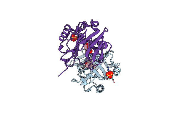 |
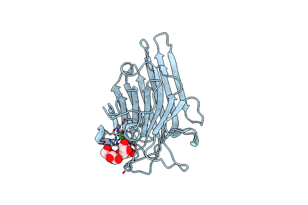
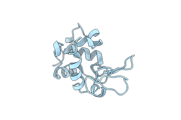
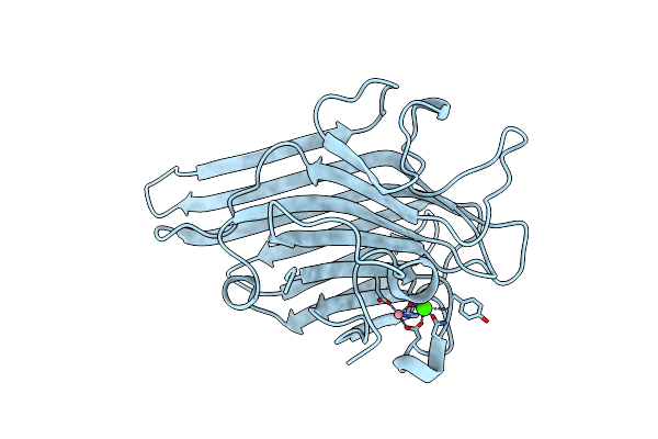
The Structure And Refinement Of Apocrustacyanin C2 To 1.6A Resolution And The Search For Differences Between This Protein And The Homologous Apoproteins A1 And C1.
Organism: Homarus gammarus
Method: X-RAY DIFFRACTION Resolution:1.60 Å Release Date: 2004-04-27 Classification: PROTEIN BINDING Ligands: SO4, MPD, GOL |
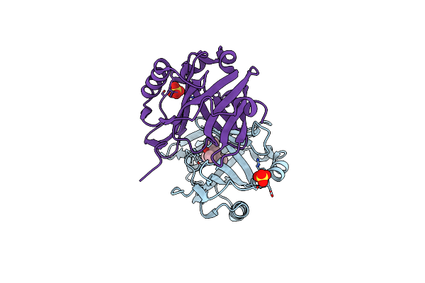 |
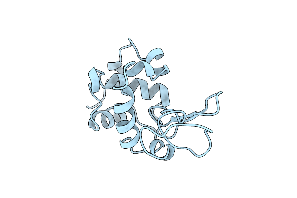
The Structure And Refinement Of Apocrustacyanin C2 To 1.3A Resolution And The Search For Differences Between This Protein And The Homologous Apoproteins A1 And C1
Organism: Homarus gammarus
Method: X-RAY DIFFRACTION Resolution:1.30 Å Release Date: 2004-03-02 Classification: PROTEIN BINDING Ligands: SO4, MPD |
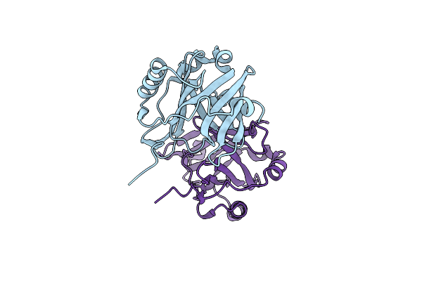 |
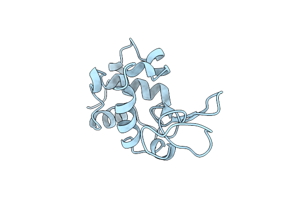
Apocrustacyanin C1 Crystals Grown In Space And Earth Using Vapour Diffusion Geometry
Organism: Homarus gammarus
Method: X-RAY DIFFRACTION Resolution:1.85 Å Release Date: 2003-07-03 Classification: TRANSPORT PROTEIN |
 |
Apocrustacyanin C1 Crystals Grown In Space And Earth Using Vapour Diffusion Geometry
Organism: Homarus gammarus
Method: X-RAY DIFFRACTION Resolution:2.00 Å Release Date: 2003-07-03 Classification: TRANSPORT PROTEIN |
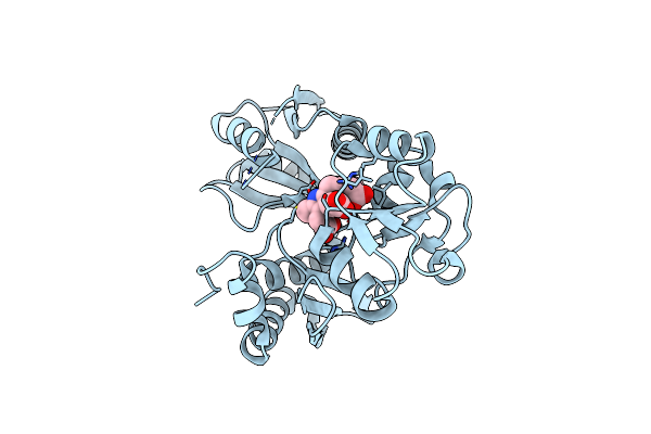 |
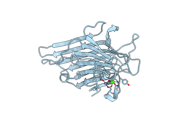
Time-Resolved And Static-Ensemble Structural Chemistry Of Hydroxymethylbilane Synthase
Organism: Escherichia coli
Method: X-RAY DIFFRACTION Resolution:1.66 Å Release Date: 2003-01-16 Classification: TRANSFERASE Ligands: DPM |
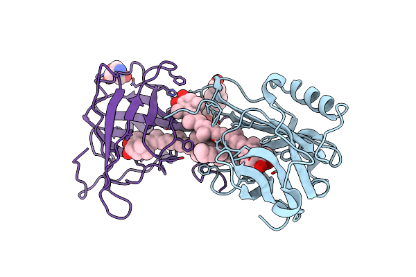 |
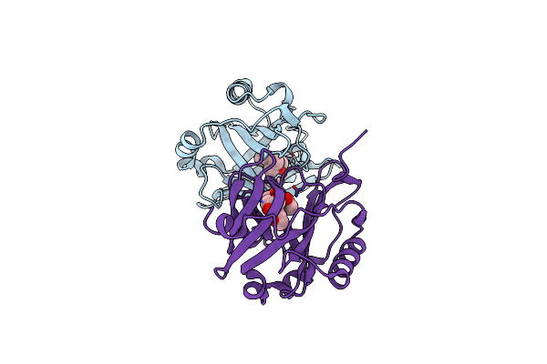
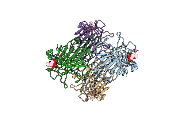
The Molecular Basis Of The Coloration Mechanism In Lobster Shell. Beta-Crustacyanin At 3.2 A Resolution
Organism: Homarus gammarus
Method: X-RAY DIFFRACTION Resolution:3.23 Å Release Date: 2002-08-08 Classification: LIPOCALIN Ligands: AXT, D12, TRS, EPE |
 |
Organism: Homarus gammarus
Method: X-RAY DIFFRACTION Resolution:1.40 Å Release Date: 2001-09-06 Classification: APOCRUSTACYANIN Ligands: MPD |
 |
Organism: Canavalia ensiformis
Method: X-RAY DIFFRACTION Resolution:1.20 Å Release Date: 2001-07-25 Classification: SUGAR BINDING PROTEIN Ligands: MN, CA |
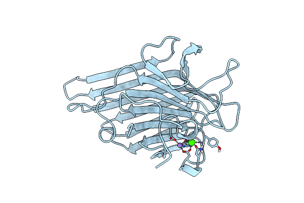 |
Direct Determination Of The Positions Of Deuterium Atoms Of Bound Water In Concanavalin A By Neutron Laue Crystallography
Organism: Canavalia ensiformis
Method: NEUTRON DIFFRACTION Resolution:2.40 Å Release Date: 2000-05-08 Classification: SUGAR BINDING PROTEIN Ligands: MN, CA |
 |
Organism: Canavalia ensiformis
Method: X-RAY DIFFRACTION Resolution:1.80 Å Release Date: 2000-04-30 Classification: LECTIN Ligands: MN, CA |
 |
Organism: Escherichia coli
Method: X-RAY DIFFRACTION Resolution:2.30 Å Release Date: 1999-02-02 Classification: TRANSFERASE Ligands: DPM |
 |
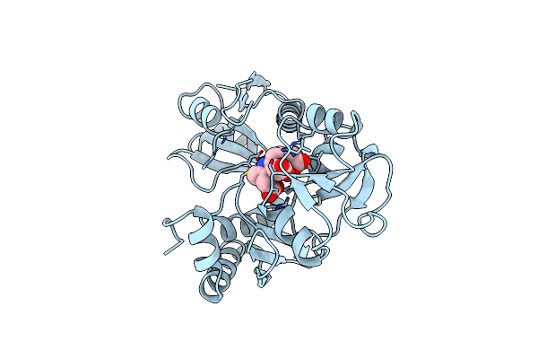
The 1.8 A Structure Of Ground Control Grown Tetragonal Hen Egg White Lysozyme
Organism: Gallus gallus
Method: X-RAY DIFFRACTION Resolution:1.80 Å Release Date: 1998-09-30 Classification: HYDROLASE |
 |
The 1.8 A Structure Of Microbatch Oil Drop Grown Tetragonal Hen Egg White Lysozyme
Organism: Gallus gallus
Method: X-RAY DIFFRACTION Resolution:1.80 Å Release Date: 1998-09-30 Classification: HYDROLASE |
 |
The 1.8 A Structure Of Microgravity Grown Tetragonal Hen Egg White Lysozyme
Organism: Gallus gallus
Method: X-RAY DIFFRACTION Resolution:1.80 Å Release Date: 1998-09-30 Classification: HYDROLASE |
 |
Organism: Gallus gallus
Method: X-RAY DIFFRACTION Resolution:1.80 Å Release Date: 1998-09-23 Classification: HYDROLASE |
 |
Organism: Canavalia ensiformis
Method: X-RAY DIFFRACTION Resolution:0.94 Å Release Date: 1997-11-26 Classification: AGGLUTININ Ligands: MN, CA |
 |
Reduced Form Selenomethionine-Labelled Hydroxymethylbilane Synthase Determined By Mad
Organism: Escherichia coli
Method: X-RAY DIFFRACTION Resolution:2.40 Å Release Date: 1997-10-15 Classification: TRANSFERASE Ligands: DPM |
 |
High-Resolution Structures Of Single-Metal-Substituted Concanavalin A: The Co,Ca-Protein At 1.6 Angstroms And The Ni,Ca-Protein At 2.0 Angstroms
Organism: Canavalia ensiformis
Method: X-RAY DIFFRACTION Resolution:2.00 Å Release Date: 1994-05-31 Classification: LECTIN(AGGLUTININ) Ligands: NI, CA |
 |
High-Resolution Structures Of Single-Metal-Substituted Concanavalin A: The Co,Ca-Protein At 1.6 Angstroms And The Ni,Ca-Protein At 2.0 Angstroms
Organism: Canavalia ensiformis
Method: X-RAY DIFFRACTION Resolution:1.60 Å Release Date: 1994-05-31 Classification: LECTIN(AGGLUTININ) Ligands: CO, CA |
 |
Refined Structure Of Concanavalin A Complexed With Alpha-Methyl-D-Mannopyranoside At 2.0 Angstroms Resolution And Comparison With The Saccharide-Free Structure
Organism: Canavalia ensiformis
Method: X-RAY DIFFRACTION Resolution:2.00 Å Release Date: 1994-05-31 Classification: LECTIN(AGGLUTININ) Ligands: MMA, MN, CA, CL |