
Search Count: 17
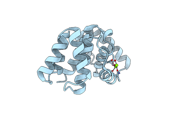 |
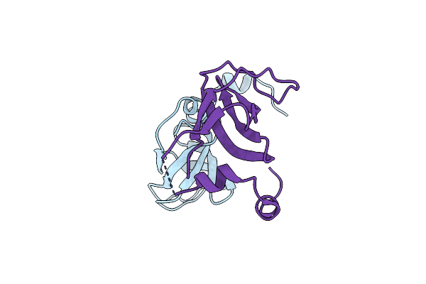
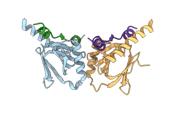
Crystal Structure Of Matrix Protein 1 From Influenza C Virus (Strain C/Ann Arbor/1/1950)
Organism: Influenza c virus (strain c/ann arbor/1/1950)
Method: X-RAY DIFFRACTION Resolution:1.50 Å Release Date: 2017-02-08 Classification: VIRAL PROTEIN Ligands: MG |
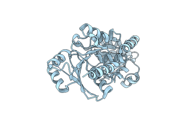 |
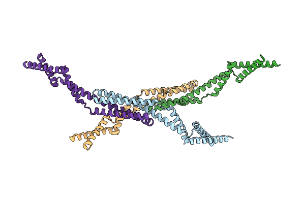
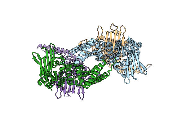
The Crystal Structure Of Arabidopsis Thaliana Galactolipid Synthase, Mgd1 (Apo-Form)
Organism: Arabidopsis thaliana
Method: X-RAY DIFFRACTION Resolution:2.50 Å Release Date: 2016-02-17 Classification: TRANSFERASE |
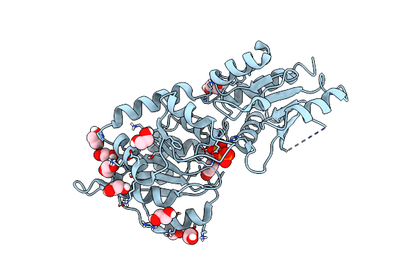 |
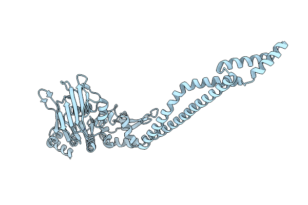
The Crystal Structure Of Arabidopsis Thaliana Galactolipid Synthase Mgd1 In Complex With Udp
Organism: Arabidopsis thaliana
Method: X-RAY DIFFRACTION Resolution:2.25 Å Release Date: 2016-02-10 Classification: TRANSFERASE Ligands: UDP, EDO |
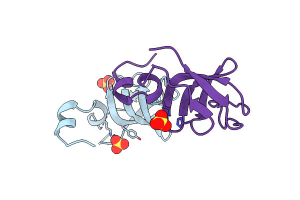 |
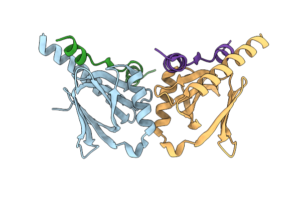
Organism: Homo sapiens
Method: X-RAY DIFFRACTION Resolution:1.87 Å Release Date: 2015-11-04 Classification: PROTEIN BINDING Ligands: SO4 |
 |
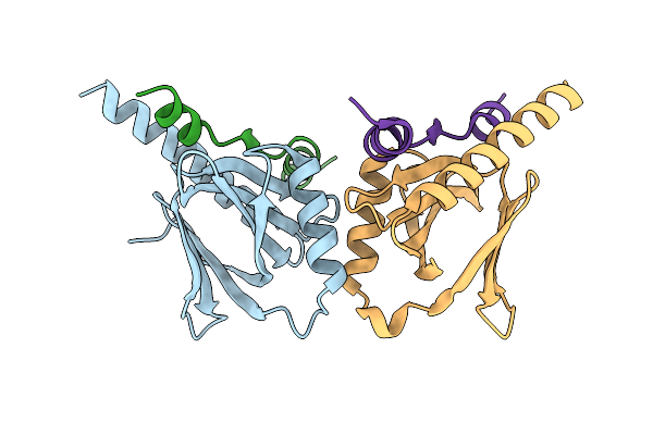
Organism: Homo sapiens
Method: X-RAY DIFFRACTION Resolution:1.73 Å Release Date: 2015-11-04 Classification: PROTEIN BINDING |
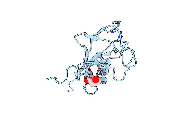 |
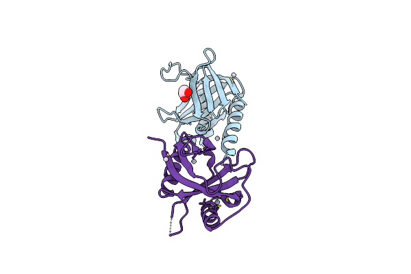
Crystal Structure Of The N-Terminal, Growth Factor-Like Domain Of The Amyloid Precursor Protein Bound To Copper
Organism: Homo sapiens
Method: X-RAY DIFFRACTION Resolution:1.75 Å Release Date: 2014-07-23 Classification: METAL BINDING PROTEIN Ligands: CU, GOL |
 |
Organism: Deinococcus radiodurans
Method: X-RAY DIFFRACTION Resolution:3.34 Å Release Date: 2013-08-07 Classification: RECOMBINATION Ligands: ZN |
 |
Organism: Deinococcus radiodurans
Method: X-RAY DIFFRACTION Resolution:2.04 Å Release Date: 2012-12-12 Classification: DNA BINDING PROTEIN |
 |
Organism: Deinococcus radiodurans
Method: X-RAY DIFFRACTION Resolution:3.00 Å Release Date: 2012-12-12 Classification: HYDROLASE |
 |
Organism: Deinococcus radiodurans
Method: X-RAY DIFFRACTION Resolution:4.00 Å Release Date: 2012-12-12 Classification: DNA BINDING PROTEIN |
 |
Organism: Arabidopsis thaliana
Method: X-RAY DIFFRACTION Resolution:1.95 Å Release Date: 2010-04-07 Classification: PLANT PROTEIN Ligands: SO4, EDO |
 |
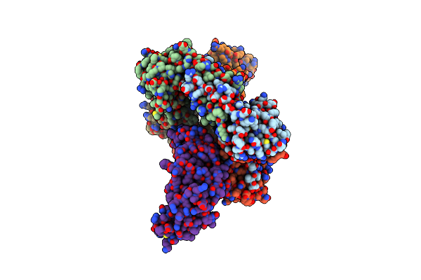
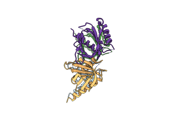
Crystal Structure Of The Intracellular Domain Of Human App In Complex With Fe65-Ptb2
Organism: Homo sapiens
Method: X-RAY DIFFRACTION Resolution:2.10 Å Release Date: 2008-09-16 Classification: PROTEIN BINDING |
 |
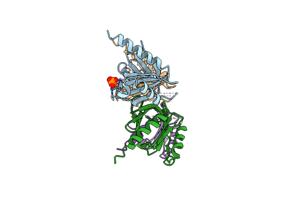
Crystal Structure Of The Intracellular Domain Of Human App (T668E Mutant) In Complex With Fe65-Ptb2
Organism: Homo sapiens
Method: X-RAY DIFFRACTION Resolution:2.20 Å Release Date: 2008-09-16 Classification: PROTEIN BINDING |
 |
Crystal Structure Of The Intracellular Domain Of Human App (T668A Mutant) In Complex With Fe65-Ptb2
Organism: Homo sapiens
Method: X-RAY DIFFRACTION Resolution:2.00 Å Release Date: 2008-09-16 Classification: PROTEIN BINDING |
 |
Organism: Homo sapiens
Method: X-RAY DIFFRACTION Resolution:2.20 Å Release Date: 2008-06-10 Classification: PROTEIN BINDING Ligands: HG, EDO |
 |
Organism: Homo sapiens
Method: X-RAY DIFFRACTION Resolution:2.80 Å Release Date: 2008-06-10 Classification: PROTEIN BINDING |
 |
Crystal Structure Of The Human Fe65-Ptb1 Domain With Bound Phosphate (Trigonal Crystal Form)
Organism: Homo sapiens
Method: X-RAY DIFFRACTION Resolution:2.70 Å Release Date: 2008-06-10 Classification: PROTEIN BINDING Ligands: PO4 |