
Search Count: 14
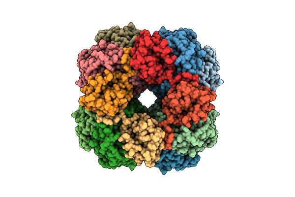 |
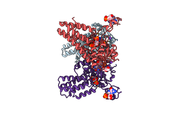
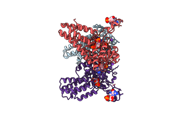
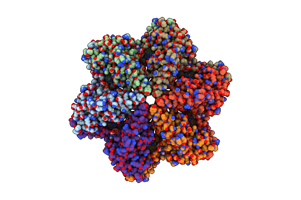
Structure Of The Far-Red Light-Absorbing Allophycocyanin Core Expressed During Farlip
Organism: Synechococcus sp. pcc 7335
Method: ELECTRON MICROSCOPY Release Date: 2024-01-03 Classification: PHOTOSYNTHESIS Ligands: RUB, MG |
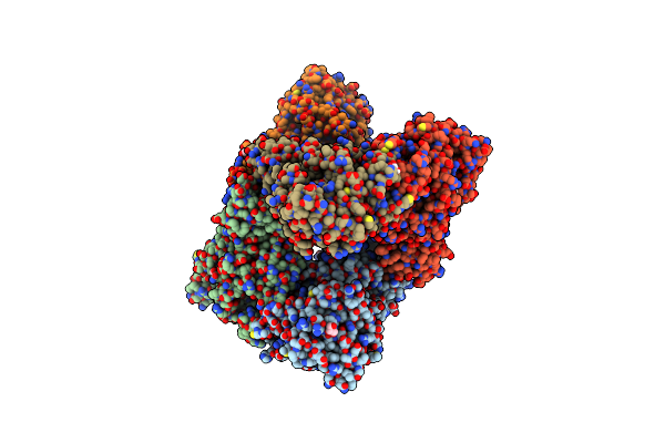 |
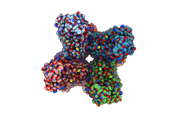
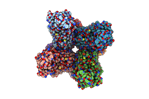
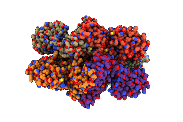
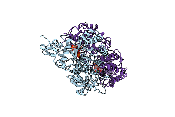
Organism: Cyanobium sp. pcc 7001
Method: X-RAY DIFFRACTION Resolution:2.30 Å Release Date: 2023-08-09 Classification: PHOTOSYNTHESIS Ligands: ZN, GOL, SO4, EDO, RUB, CO2, BCT |
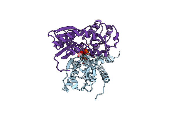 |
Crystal Structure Of The Effector-Binding Domain Of Synechococcus Elongatus Cmpr In Complex With Ribulose-1,5-Bisphosphate
Organism: Synechococcus elongatus pcc 7942
Method: X-RAY DIFFRACTION Resolution:2.15 Å Release Date: 2018-10-10 Classification: TRANSCRIPTION Ligands: RUB |
 |
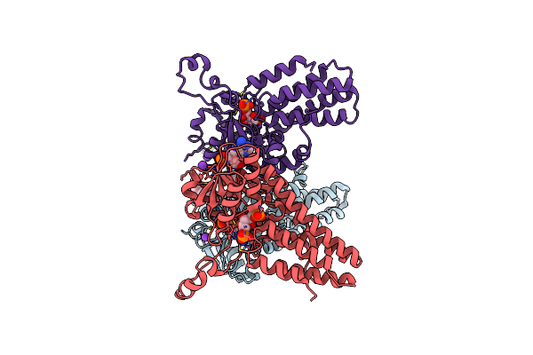
Crystal Structure Of Ribose-1,5-Bisphosphate Isomerase From Pyrococcus Horikoshii Ot3 In Complex With Ribulose-1,5-Bisphosphate
Organism: Pyrococcus horikoshii
Method: X-RAY DIFFRACTION Resolution:2.20 Å Release Date: 2018-02-14 Classification: ISOMERASE Ligands: RUB, K, CL, PEG, MPD |
 |
Crystal Structure Of Ribose-1,5-Bisphosphate Isomerase From Pyrococcus Horikoshii Ot3 In Complex With Ribulose-1,5-Bisphosphate And Amp
Organism: Pyrococcus horikoshii
Method: X-RAY DIFFRACTION Resolution:2.35 Å Release Date: 2018-02-14 Classification: ISOMERASE Ligands: RUB, K, AMP, MPD |
 |
Crystal Structure Of Ribose-1,5-Bisphosphate Isomerase From Pyrococcus Horikoshii Ot3 In Complex With Ribulose-1,5-Bisphosphate And Gmp
Organism: Pyrococcus horikoshii
Method: X-RAY DIFFRACTION Resolution:2.75 Å Release Date: 2018-02-14 Classification: ISOMERASE Ligands: RUB, MPD, K, PEG, 5GP |
 |
Crystal Structure Of Ribose-1,5-Bisphosphate Isomerase From Pyrococcus Horikoshii Ot3 In Complex With Ribulose-1,5-Bisphosphate, Amp And Gmp
Organism: Pyrococcus horikoshii
Method: X-RAY DIFFRACTION Resolution:2.80 Å Release Date: 2018-02-14 Classification: ISOMERASE Ligands: RUB, MPD, AMP, K, 5GP |
 |
Organism: Pisum sativum
Method: X-RAY DIFFRACTION Resolution:2.15 Å Release Date: 2013-10-16 Classification: LYASE Ligands: RUB, PO4, A8S |
 |
Organism: Pisum sativum
Method: X-RAY DIFFRACTION Resolution:2.20 Å Release Date: 2012-10-31 Classification: LYASE Ligands: RUB |
 |
Negative Stain Em Map Of The Aaa Protein Cbbx, A Red-Type Rubisco Activase From R. Sphaeroides
Organism: Rhodobacter sphaeroides
Method: ELECTRON MICROSCOPY Resolution:21.00 Å Release Date: 2011-11-09 Classification: ATP BINDING PROTEIN Ligands: RUB, ADP |
 |
Crystal Structure Of Ribose-1,5-Bisphosphate Isomerase From Thermococcus Kodakaraensis Kod1 In Complex With Ribulose-1,5-Bisphosphate
Organism: Thermococcus kodakarensis
Method: X-RAY DIFFRACTION Resolution:2.60 Å Release Date: 2010-11-03 Classification: ISOMERASE Ligands: RUB, PEG, MG |
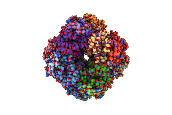 |
Non-Activated Spinach Rubisco In Complex With Its Substrate Ribulose-1,5-Bisphosphate
Organism: Spinacia oleracea
Method: X-RAY DIFFRACTION Resolution:2.40 Å Release Date: 1997-06-16 Classification: LYASE (CARBON-CARBON) Ligands: RUB |
 |
Activated Spinach Rubisco In Complex With Its Substrate Ribulose-1,5-Bisphosphate And Calcium
Organism: Spinacia oleracea
Method: X-RAY DIFFRACTION Resolution:2.20 Å Release Date: 1997-06-16 Classification: LYASE (CARBON-CARBON) Ligands: RUB, CA |
 |
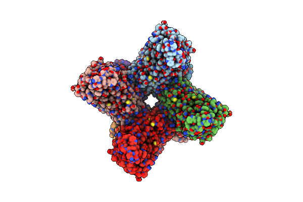
Crystal Structure Of Activated Ribulose-1,5-Bisphosphate Carboxylase Complexed With Its Substrate, Ribulose-1,5-Bisphosphate
Organism: Rhodospirillum rubrum
Method: X-RAY DIFFRACTION Resolution:2.60 Å Release Date: 1993-01-15 Classification: LYASE(CARBON-CARBON) Ligands: RUB, MG, FMT |