
Search Count: 36,883
 |
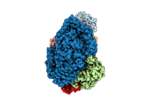
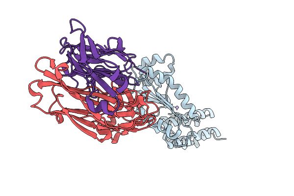
Fadd-Ded Filaments Coordinate Complex Iia Assembly During Tnf-Induced Apoptosis
Organism: Homo sapiens
Method: ELECTRON MICROSCOPY Release Date: 2025-12-31 Classification: IMMUNE SYSTEM |
 |
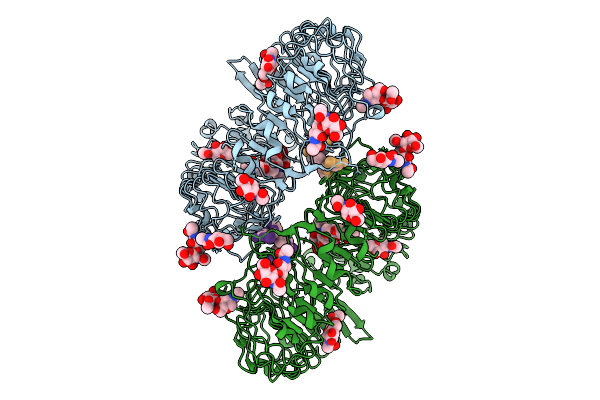
Organism: Archaeoglobus fulgidus dsm 4304, Pyrococcus furiosus dsm 3638
Method: ELECTRON MICROSCOPY Release Date: 2025-12-31 Classification: ANTIVIRAL PROTEIN Ligands: MG, ATP, ZN |
 |
Organism: Archaeoglobus fulgidus dsm 4304, Archaeoglobus fulgidus, Pyrococcus furiosus dsm 3638
Method: ELECTRON MICROSCOPY Release Date: 2025-12-31 Classification: ANTIVIRAL PROTEIN Ligands: MG, ATP, ZN |
 |
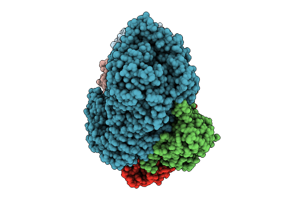
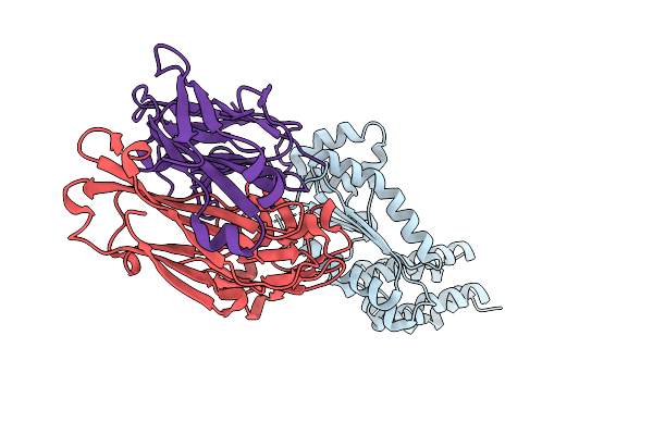
Organism: Macaca mulatta, Homo sapiens
Method: ELECTRON MICROSCOPY Release Date: 2025-12-31 Classification: IMMUNE SYSTEM Ligands: NAG |
 |
Organism: Homo sapiens
Method: X-RAY DIFFRACTION Release Date: 2025-12-31 Classification: RNA BINDING PROTEIN |
 |
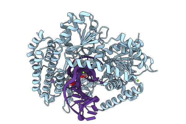
Structure Of Chicken Lgp2 Bound To 10-Mer Rna Mismatched Duplex That Mimics The Influenza B Virus Vrna Promoter (Panhandle) And To Adp-Alf4-Mg.
Organism: Gallus gallus, Influenza b virus
Method: X-RAY DIFFRACTION Release Date: 2025-12-31 Classification: RNA BINDING PROTEIN Ligands: ZN, ADP, ALF, MG |
 |
Structure Of Duck Rig-I (Delta Cards) Bound To 31-Mer Rna Mismatched Hairpin With 5'Pppg-C Blunt End, Mimicking The Influenza B Virus Vrna Promoter (Panhandle).
Organism: Anas platyrhynchos, Influenza b virus
Method: X-RAY DIFFRACTION Release Date: 2025-12-31 Classification: RNA BINDING PROTEIN Ligands: ZN, MG, CL, K, GOL, GTP |
 |
Structure Of Duck Rig-I (Delta Cards) Bound To 31-Mer Rna Mismatched Hairpin With 5'Ppa-U Blunt End Mimicking The Influenza B Virus Vrna Promoter (Panhandle) And To Adp.
Organism: Anas platyrhynchos, Influenza b virus
Method: X-RAY DIFFRACTION Release Date: 2025-12-31 Classification: RNA BINDING PROTEIN Ligands: ZN, ADP, GOL, K, MG |
 |
Structure Of Duck Rig-I (Delta Cards) Bound To 31-Mer Rna Mismatched Hairpin With 5'Pppg-C Blunt End Mimicking The Influenza A Virus Vrna Promoter (Panhandle) And To Adp.
Organism: Anas platyrhynchos, Influenza a virus
Method: X-RAY DIFFRACTION Release Date: 2025-12-31 Classification: RNA BINDING PROTEIN Ligands: ZN, ADP, CL, MG, GTP |
 |
Crystal Structure Of Crimean-Congo Hemorrhagic Fever Virus Cap-Snatching Endonuclease
Organism: Orthonairovirus haemorrhagiae, Mus musculus
Method: X-RAY DIFFRACTION Release Date: 2025-12-31 Classification: HYDROLASE |
 |
Organism: Kasokero virus, Mus musculus
Method: X-RAY DIFFRACTION Release Date: 2025-12-31 Classification: HYDROLASE |
 |
Crystal Structure Of Kasokero Virus Cap- Snatching Endonuclease In Complex With Manganese Ions
Organism: Kasokero virus, Mus musculus
Method: X-RAY DIFFRACTION Release Date: 2025-12-31 Classification: HYDROLASE Ligands: MN |
 |
Crystal Structure Of Kasokero Virus Cap- Snatching Endonuclease (E668A Mutant)
Organism: Kasokero virus, Mus musculus
Method: X-RAY DIFFRACTION Release Date: 2025-12-31 Classification: HYDROLASE Ligands: MN |
 |
Crystal Structure Of Kasokero Virus Cap- Snatching Endonuclease (E682A Mutant)
Organism: Kasokero virus, Mus musculus
Method: X-RAY DIFFRACTION Release Date: 2025-12-31 Classification: HYDROLASE Ligands: MN |
 |
Crystal Structure Of Kasokero Virus Cap- Snatching Endonuclease In Complex With 2,4-Dioxo-4-Phenylbutanoic Acid (Dpba)
Organism: Kasokero virus, Mus musculus
Method: X-RAY DIFFRACTION Release Date: 2025-12-31 Classification: HYDROLASE Ligands: MN, XI7 |
 |
Crystal Structure Of Kasokero Virus Cap- Snatching Endonuclease In Complex With L-742,001
Organism: Kasokero virus, Mus musculus
Method: X-RAY DIFFRACTION Release Date: 2025-12-31 Classification: HYDROLASE Ligands: MN, 0N8 |
 |
Crystal Structure Of Kasokero Virus Cap- Snatching Endonuclease In Complex With Baloxavir Acid (Bxa)
Organism: Kasokero virus, Mus musculus
Method: X-RAY DIFFRACTION Release Date: 2025-12-31 Classification: HYDROLASE Ligands: MN, E4Z |
 |
Organism: Escherichia coli
Method: ELECTRON MICROSCOPY Release Date: 2025-12-31 Classification: DNA BINDING PROTEIN/RNA/DNA Ligands: ATP, MG |
 |
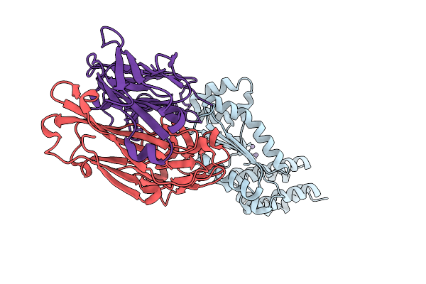
Organism: Streptococcus pyogenes serotype m1, Synthetic construct
Method: ELECTRON MICROSCOPY Release Date: 2025-12-31 Classification: IMMUNE SYSTEM |
 |
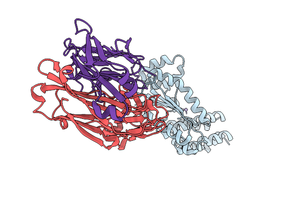
Organism: Streptococcus pyogenes serotype m1, Synthetic construct
Method: ELECTRON MICROSCOPY Release Date: 2025-12-31 Classification: IMMUNE SYSTEM |