
Search Count: 1,289
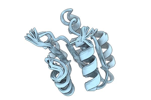 |
Organism: Thermotoga maritima msb8
Method: SOLUTION NMR Release Date: 2026-01-14 Classification: SIGNALING PROTEIN |
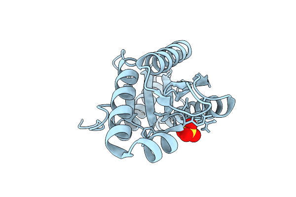 |
Organism: Pseudomonas aeruginosa
Method: X-RAY DIFFRACTION Release Date: 2025-12-10 Classification: SIGNALING PROTEIN Ligands: SO4 |
 |
Structure Of The Acinetobacter Baumannii Response Regulator Pmra Receiver Domain D10N Mutation
Organism: Acinetobacter baumannii
Method: X-RAY DIFFRACTION Release Date: 2025-11-26 Classification: TRANSCRIPTION Ligands: MG |
 |
Structure Of The Acinetobacter Baumannii Response Regulator Pmra Receiver Domain M12I Mutation
Organism: Acinetobacter baumannii
Method: X-RAY DIFFRACTION Release Date: 2025-11-26 Classification: TRANSCRIPTION Ligands: MG |
 |
Structure Of The Acinetobacter Baumannii Response Regulator Pmra Receiver Domain I13M Mutation
Organism: Acinetobacter baumannii
Method: X-RAY DIFFRACTION Release Date: 2025-11-26 Classification: TRANSCRIPTION Ligands: MG |
 |
Structure Of The Acinetobacter Baumannii Response Regulator Pmra Receiver Domain I13M Mutation In Active Dimer State
Organism: Acinetobacter baumannii
Method: X-RAY DIFFRACTION Release Date: 2025-11-26 Classification: TRANSCRIPTION |
 |
Structure Of The Acinetobacter Baumannii Response Regulator Pmra Receiver Domain G54E Mutation
Organism: Acinetobacter baumannii
Method: X-RAY DIFFRACTION Release Date: 2025-11-26 Classification: TRANSCRIPTION Ligands: MG |
 |
Structure Of The Acinetobacter Baumannii Response Regulator Pmra Receiver Domain S119T Mutation
Organism: Acinetobacter baumannii
Method: X-RAY DIFFRACTION Release Date: 2025-11-26 Classification: TRANSCRIPTION |
 |
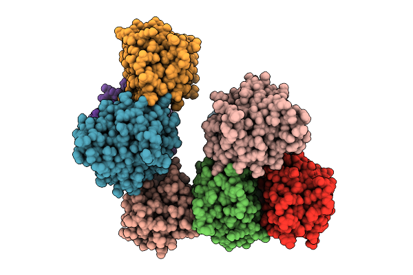
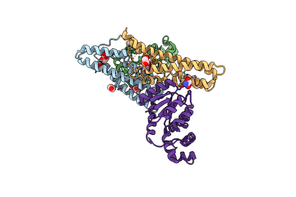
Cryoem Structure Of Rpaa Bound To Pkaibc Dna And Syn7942 Rnap-Siga Holoenzyme
Organism: Synechococcus elongatus, Synechococcus elongatus pcc 7942 = fachb-805, Synthetic construct
Method: ELECTRON MICROSCOPY Release Date: 2025-10-15 Classification: DNA BINDING PROTEIN/DNA Ligands: MG, ZN |
 |
Organism: Synthetic construct
Method: X-RAY DIFFRACTION Release Date: 2025-10-01 Classification: DE NOVO PROTEIN |
 |
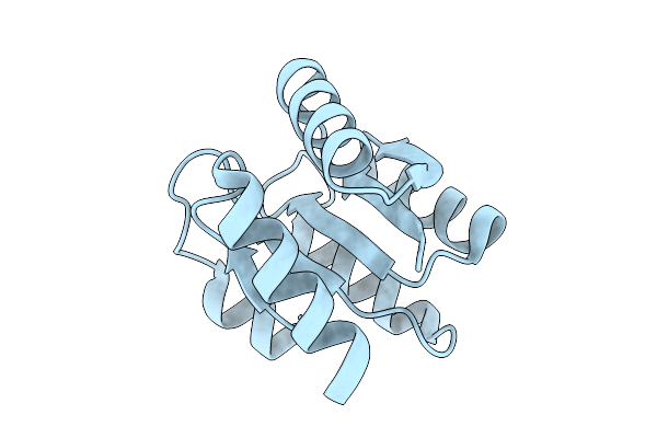
Solution Structure Of Staphylococcus Aureus Response Regulator Arlr Dna-Binding Domain
Organism: Staphylococcus aureus subsp. aureus nctc 8325
Method: SOLUTION NMR Release Date: 2025-09-17 Classification: DNA BINDING PROTEIN |
 |
Organism: Caulobacter vibrioides
Method: X-RAY DIFFRACTION Release Date: 2025-08-06 Classification: SIGNALING PROTEIN Ligands: C2E, G4P, MG |
 |
Organism: Caulobacter vibrioides
Method: X-RAY DIFFRACTION Release Date: 2025-08-06 Classification: SIGNALING PROTEIN Ligands: C2E, 0O2, MG |
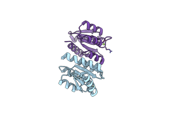 |
The Rec Domain (In The Non-Phosphorylated State) Of Xync, A Response Regulator From G.Proteiniphilus T-6
Organism: Geobacillus proteiniphilus
Method: X-RAY DIFFRACTION Release Date: 2025-06-25 Classification: DNA BINDING PROTEIN Ligands: MG |
 |
Organism: Rubellimicrobium thermophilum dsm 16684
Method: X-RAY DIFFRACTION Release Date: 2025-05-28 Classification: SIGNALING PROTEIN |
 |
Organism: Rubellimicrobium thermophilum dsm 16684
Method: X-RAY DIFFRACTION Resolution:1.99 Å Release Date: 2025-05-21 Classification: SIGNALING PROTEIN Ligands: MG, NA, TRS |
 |
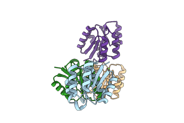
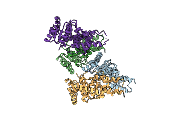
Complex Of Histidine-Containing Phosphotransfer 1 (Ahp1) And Response Regulator 1 (Arr1) From A. Thaliana
Organism: Arabidopsis thaliana
Method: X-RAY DIFFRACTION Resolution:2.87 Å Release Date: 2025-03-26 Classification: SIGNALING PROTEIN Ligands: OXM, EDO, GOL |
 |
Organism: Metschnikowia bicuspidata, Myxococcus xanthus, Homo sapiens
Method: X-RAY DIFFRACTION Release Date: 2025-03-12 Classification: CHAPERONE |
 |
Organism: Leptospira interrogans serovar copenhageni
Method: X-RAY DIFFRACTION Resolution:2.65 Å Release Date: 2024-12-18 Classification: SIGNALING PROTEIN Ligands: ACP, MG, BEF, SO4 |
 |
Organism: Homo sapiens
Method: ELECTRON MICROSCOPY Release Date: 2024-09-18 Classification: SPLICING Ligands: GTP, ZN, IHP |