Search Count: 7,711
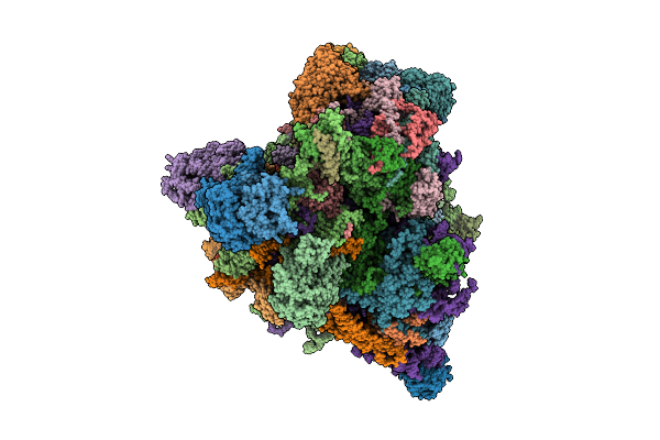 |
Translational Activator Aep3 In Complex With Mrna And The Yeast Mitochondrial Ribosome
Organism: Saccharomyces cerevisiae w303, Saccharomyces cerevisiae
Method: ELECTRON MICROSCOPY Release Date: 2025-12-17 Classification: RIBOSOME Ligands: MG, GTP |
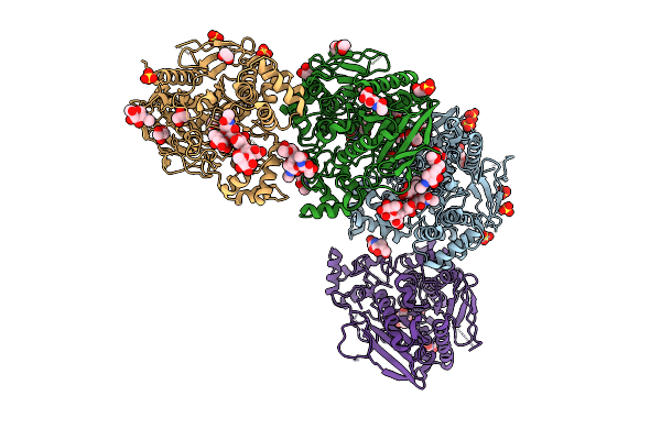 |
Organism: Sus scrofa
Method: X-RAY DIFFRACTION Release Date: 2025-12-03 Classification: HYDROLASE Ligands: NAG, GOL, PG4, SO4, PEG, EDO |
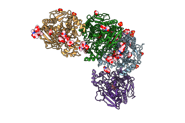 |
Organism: Sus scrofa
Method: X-RAY DIFFRACTION Release Date: 2025-12-03 Classification: HYDROLASE Ligands: NAG, GOL, GB, SO4, PEG, EDO |
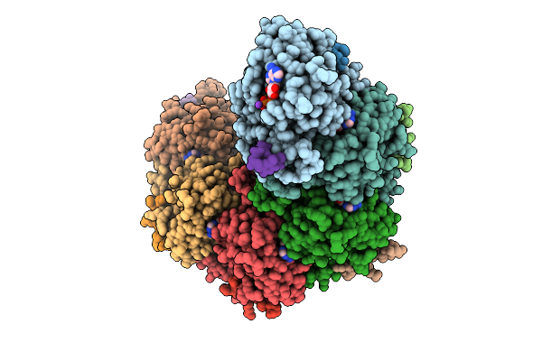 |
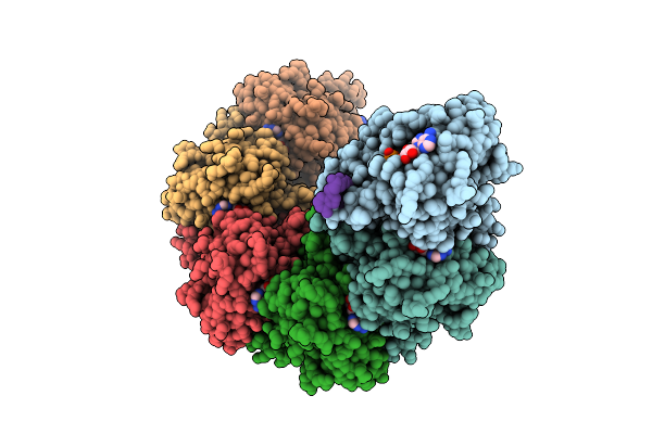
Rad51 Filament In Complex With Calcium And Atp Bound By The Rad51Ap1 C-Terminus
Organism: Homo sapiens, Synthetic construct
Method: ELECTRON MICROSCOPY Release Date: 2025-12-03 Classification: DNA BINDING PROTEIN Ligands: ATP, CA, K |
 |
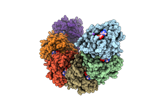
Organism: Homo sapiens, Synthetic construct
Method: ELECTRON MICROSCOPY Release Date: 2025-12-03 Classification: DNA BINDING PROTEIN Ligands: ATP, MG, K |
 |
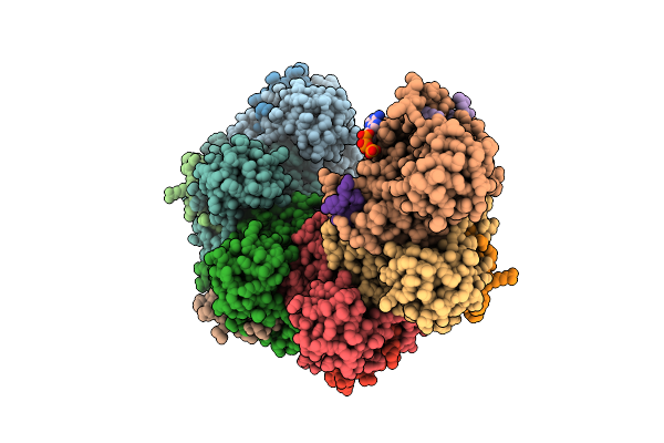
Organism: Homo sapiens
Method: ELECTRON MICROSCOPY Release Date: 2025-12-03 Classification: DNA BINDING PROTEIN Ligands: MG, ADP, K |
 |
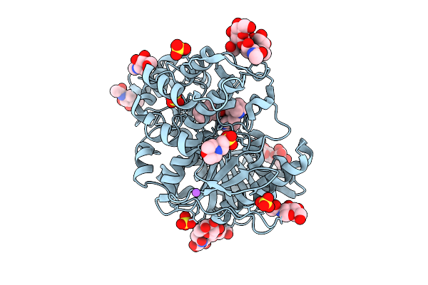
Rad51 Filament In Complex With Magnesium And Atp Bound By The Rad51Ap1 C-Terminus
Organism: Homo sapiens, Synthetic construct
Method: ELECTRON MICROSCOPY Release Date: 2025-12-03 Classification: DNA BINDING PROTEIN Ligands: ATP, K, MG |
 |
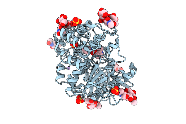
Structure Of Recombinant Human Butyrylcholinesterase In Complex With Naphthalen-2-Yl Methyl((2S,3R)-2-(Pyridin-3-Ylmethyl)Quinuclidin-3-Yl)Carbamate
Organism: Homo sapiens
Method: X-RAY DIFFRACTION Release Date: 2025-11-26 Classification: HYDROLASE Ligands: NAG, A1JOH, MES, SO4, NA |
 |
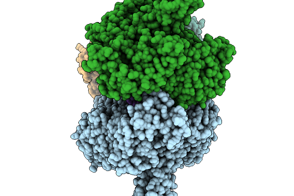
Structure Of Recombinant Human Butyrylcholinesterase In Complex With (2S, 3R)-2-(Pyridin-3-Ylmethyl)Quinuclidin-3-Yl Phenylcarbamate
Organism: Homo sapiens
Method: X-RAY DIFFRACTION Release Date: 2025-11-26 Classification: HYDROLASE Ligands: NAG, A1JOI, SO4, CL, NA |
 |
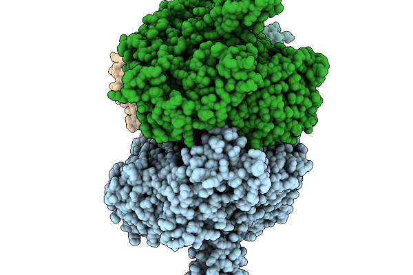
T4 Bacteriophage Replicative Polymerase Captured In Polymerase Exchange State 1
Organism: Escherichia phage t4, Synthetic construct
Method: ELECTRON MICROSCOPY Release Date: 2025-11-19 Classification: replication/DNA |
 |
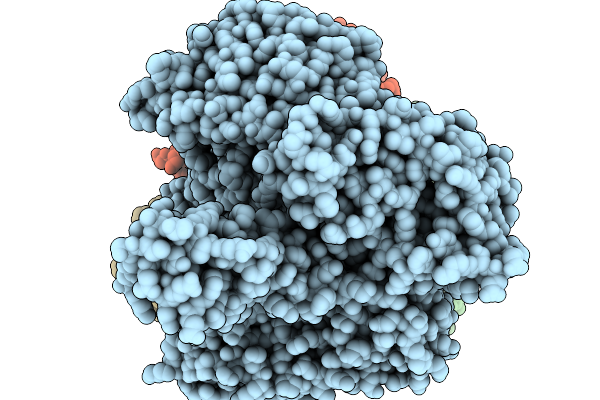
T4 Bacteriophage Replicative Polymerase Captured In Polymerase Exchange State 2
Organism: Escherichia phage t4, Synthetic construct
Method: ELECTRON MICROSCOPY Release Date: 2025-11-19 Classification: replication/DNA |
 |
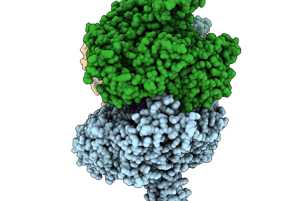
T4 Bacteriophage Replicative Holoenzyme Containing Triple Mutations D75R, Q430E, And K432E In The Exonuclease-Deficient Polymerase
Organism: Escherichia phage t4, Synthetic construct
Method: ELECTRON MICROSCOPY Release Date: 2025-11-19 Classification: Transferase/DNA |
 |
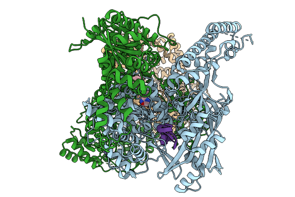
T4 Bacteriophage Replicative Polymerase Captured In Polymerase Exchange State 3
Organism: Escherichia phage t4, Synthetic construct
Method: ELECTRON MICROSCOPY Release Date: 2025-11-19 Classification: Replication/DNA Ligands: CA, D3T |
 |
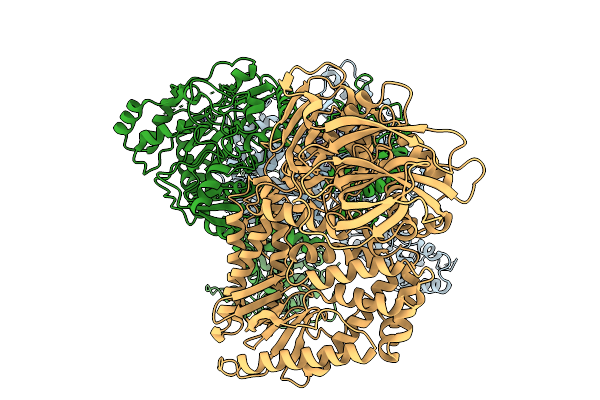
Composite Structure Of Hsv-1 Helicase-Primase In Complex With A Forked Dna And Amenamevir
Organism: Human alphaherpesvirus 1 strain 17, Homo sapiens
Method: ELECTRON MICROSCOPY Release Date: 2025-11-19 Classification: Transferase/Hydrolase Ligands: A1BXD, ZN |
 |
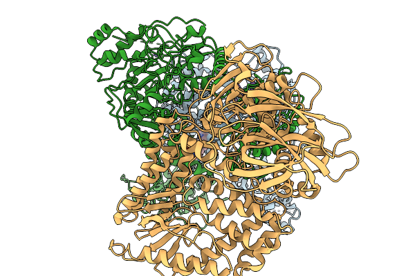
Organism: Human alphaherpesvirus 1 strain 17, Homo sapiens
Method: ELECTRON MICROSCOPY Release Date: 2025-11-19 Classification: Transferase/Hydrolase/DNA Ligands: ZN |
 |
Composite Structure Of Hsv1 Helicase-Primase In Complex With A Forked Dna And Pritelivir
Organism: Human alphaherpesvirus 1 strain 17, Homo sapiens
Method: ELECTRON MICROSCOPY Release Date: 2025-11-19 Classification: VIRAL PROTEIN/DNA Ligands: A1BXB, ZN |
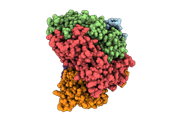 |
Structure Of The S. Cerevisiae Clamp Loader Replication Factor C (Rfc) With Mixed Nucleotide Occupancy
Organism: Saccharomyces cerevisiae
Method: ELECTRON MICROSCOPY Release Date: 2025-11-12 Classification: REPLICATION Ligands: ACT, ADP, GDP |
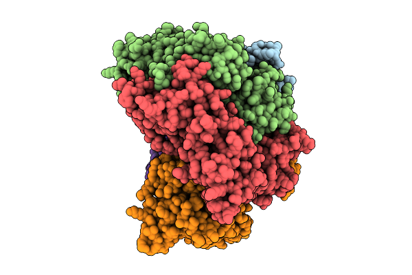 |
Structure Of The S. Cerevisiae Clamp Loader Replication Factor C (Rfc) With Mixed Nucleotide Occupancy
Organism: Saccharomyces cerevisiae
Method: ELECTRON MICROSCOPY Release Date: 2025-11-12 Classification: REPLICATION Ligands: ACT, ADP, GDP |
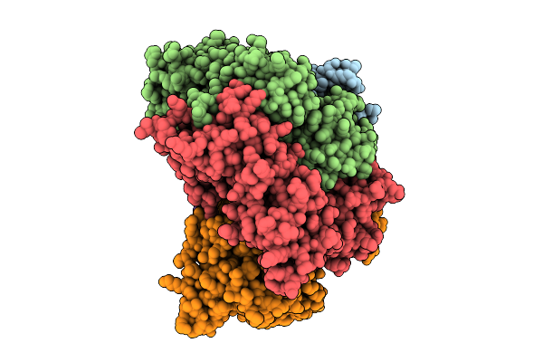 |
Structure Of The S. Cerevisiae Clamp Loader Replication Factor C (Rfc) With Mixed Nucleotide Occupancy
Organism: Saccharomyces cerevisiae
Method: ELECTRON MICROSCOPY Release Date: 2025-11-12 Classification: REPLICATION Ligands: ACT, ADP, GDP |
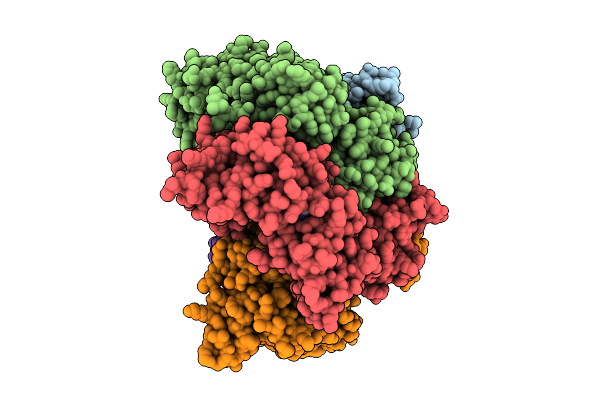 |
Structure Of The S. Cerevisiae Clamp Loader Replication Factor C (Rfc) With Mixed Nucleotide Occupancy
Organism: Saccharomyces cerevisiae
Method: ELECTRON MICROSCOPY Release Date: 2025-11-12 Classification: REPLICATION Ligands: ACT, ADP, GDP |

