
Search Count: 86
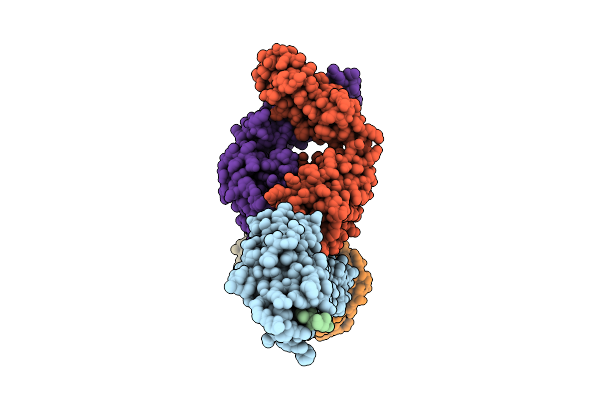 |
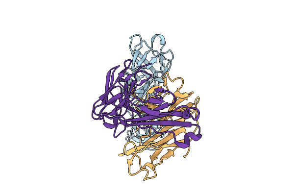
Antibody Fragments From Mab475 And Mab824 Bound To The Adhesin Protein Fimh
Organism: Escherichia coli, Escherichia coli k-12, Mus musculus
Method: ELECTRON MICROSCOPY Release Date: 2025-11-26 Classification: CELL ADHESION/IMMUNE SYSTEM |
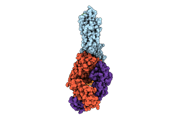 |
Antibody Fragments From Mab824 And Mab926 Bound To The Adhesin Protein Fimh
Organism: Escherichia coli, Mus musculus
Method: ELECTRON MICROSCOPY Release Date: 2025-11-26 Classification: CELL ADHESION/IMMUNE SYSTEM |
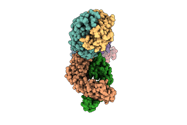 |
Antibody Fragments From Mab21, Mab475, And Mab824 Bound To The Adhesin Protein Fimh
Organism: Escherichia coli, Mus musculus
Method: ELECTRON MICROSCOPY Release Date: 2025-11-26 Classification: CELL ADHESION/IMMUNE SYSTEM |
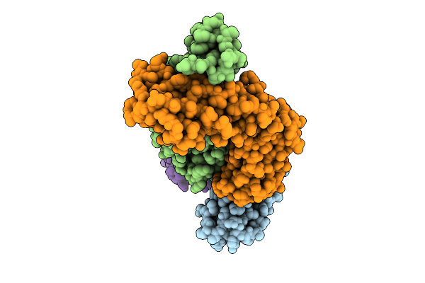 |
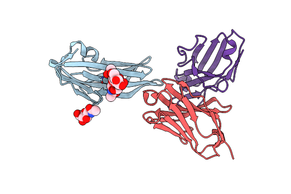
Antibody Fragments From Mab21 And Mab824 Bound To The Adhesin Protein Fimh Containing Alpha-Methyl Mannose
Organism: Escherichia coli, Mus musculus
Method: ELECTRON MICROSCOPY Release Date: 2025-11-26 Classification: CELL ADHESION/IMMUNE SYSTEM Ligands: MMA |
 |
Organism: Escherichia coli, Mus musculus
Method: ELECTRON MICROSCOPY Release Date: 2025-11-26 Classification: CELL ADHESION/IMMUNE SYSTEM |
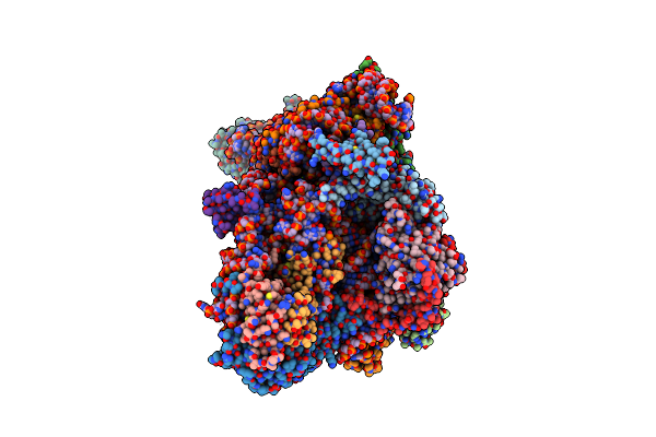 |
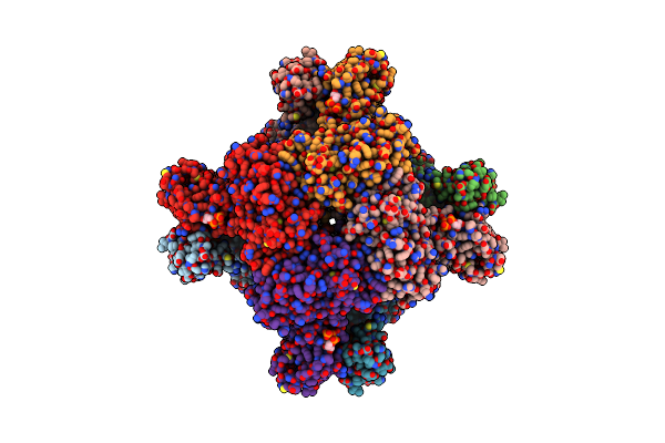
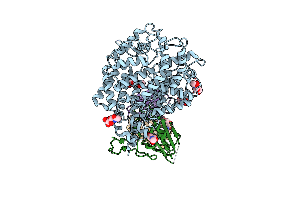
Sars-Cov-2 Nsp1 Bound To The Rhinolophus Lepidus 40S Ribosomal Subunit (Local Refinement Of The 40S Body)
Organism: Severe acute respiratory syndrome coronavirus 2, Homo sapiens, Rhinolophus lepidus
Method: ELECTRON MICROSCOPY Release Date: 2025-06-11 Classification: RIBOSOME Ligands: MG, K, ZN |
 |
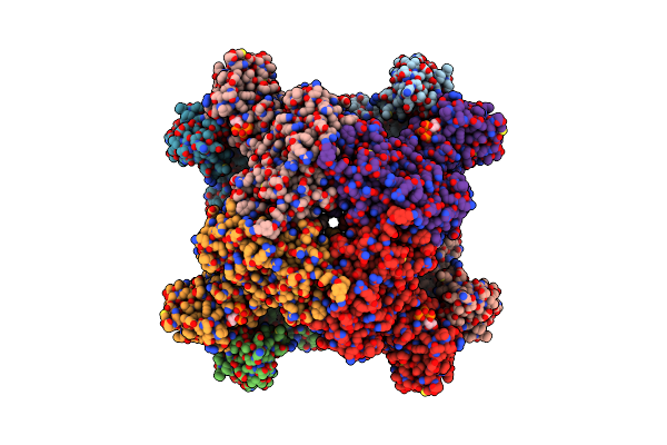
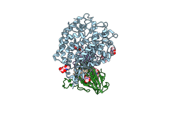
Sars-Cov-2 Nsp1 Bound To The Rhinolophus Lepidus 40S Ribosome (Local Refinement Of The 40S Head)
Organism: Rhinolophus lepidus
Method: ELECTRON MICROSCOPY Release Date: 2025-06-11 Classification: RIBOSOME Ligands: MG, ZN, K |
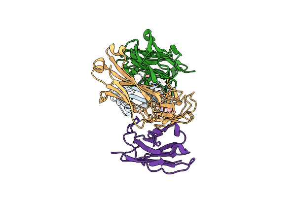 |
Structure Of The Porcine Deltacoronavirus (Pdcov) Receptor-Binding Domain Bound To The Pd33 Antibody Fab Fragment And The Kappa Light Chain Nanobody
Organism: Porcine deltacoronavirus, Mus sp., Lama glama
Method: ELECTRON MICROSCOPY Release Date: 2024-11-13 Classification: VIRAL PROTEIN/IMMUNE SYSTEM Ligands: NAG |
 |
Organism: Porcine deltacoronavirus, Mus musculus
Method: ELECTRON MICROSCOPY Release Date: 2024-11-13 Classification: VIRAL PROTEIN Ligands: NAG, PAM |
 |
Organism: Porcine deltacoronavirus, Mus musculus
Method: ELECTRON MICROSCOPY Release Date: 2024-11-13 Classification: VIRAL PROTEIN Ligands: NAG |
 |
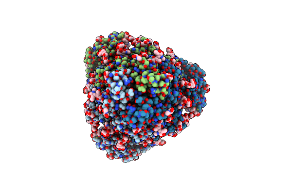
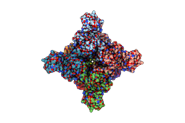
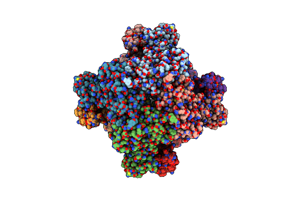
Human Retinal Variant Phosphomimetic Impdh1(595)-S477D Free Octamer Bound By Gtp, Atp, Imp, And Nad+
Organism: Homo sapiens
Method: ELECTRON MICROSCOPY Release Date: 2024-01-31 Classification: OXIDOREDUCTASE Ligands: GTP, ATP, IMP, NAD |
 |
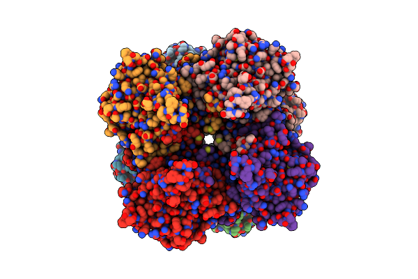
Human Retinal Variant Phosphomimetic Impdh1(546)-S477D Filament Bound By Gtp, Atp, Imp, And Nad+, Octamer-Centered
Organism: Homo sapiens
Method: ELECTRON MICROSCOPY Release Date: 2024-01-31 Classification: OXIDOREDUCTASE Ligands: GTP, NAD, IMP, ATP |
 |
Human Retinal Variant Phosphomimetic Impdh1(546)-S477D Filament Bound By Gtp, Atp, Imp, And Nad+, Interface-Centered
Organism: Homo sapiens
Method: ELECTRON MICROSCOPY Release Date: 2024-01-31 Classification: OXIDOREDUCTASE Ligands: GTP, NAD, IMP, ATP |
 |
Human Retinal Variant Phosphomimetic Impdh1(546)-S477D Filament Bound By Atp, Imp, And Nad+, Octamer-Centered
Organism: Homo sapiens
Method: ELECTRON MICROSCOPY Release Date: 2024-01-31 Classification: OXIDOREDUCTASE Ligands: ATP, IMP, NAD |
 |
Human Retinal Variant Phosphomimetic Impdh1(546)-S477D Filament Bound By Atp, Imp, And Nad+, Interface-Centered
Organism: Homo sapiens
Method: ELECTRON MICROSCOPY Release Date: 2024-01-31 Classification: OXIDOREDUCTASE Ligands: IMP, NAD |
 |
Organism: Rhinolophus alcyone, Sarbecovirus
Method: ELECTRON MICROSCOPY Release Date: 2023-12-06 Classification: VIRAL PROTEIN Ligands: NAG, ZN |
 |
Organism: Sarbecovirus sp.
Method: ELECTRON MICROSCOPY Release Date: 2023-12-06 Classification: VIRAL PROTEIN Ligands: NAG |
 |
Sars-Cov-2 Xbb.1 Spike Rbd Bound To The Human Ace2 Ectodomain And The S309 Neutralizing Antibody Fab Fragment
Organism: Homo sapiens, Severe acute respiratory syndrome coronavirus
Method: ELECTRON MICROSCOPY Release Date: 2023-10-04 Classification: VIRAL PROTEIN/IMMUNE SYSTEM Ligands: NAG |
 |
Sars-Cov-2 Bq.1.1 Spike Rbd Bound To The Human Ace2 Ectodomain And The S309 Neutralizing Antibody Fab Fragment
Organism: Homo sapiens, Severe acute respiratory syndrome coronavirus 2
Method: ELECTRON MICROSCOPY Release Date: 2023-10-04 Classification: VIRAL PROTEIN/IMMUNE SYSTEM Ligands: NAG, ZN |
 |
Sars-Cov-2 Bn.1 Spike Rbd Bound To The Human Ace2 Ectodomain And The S309 Neutralizing Antibody Fab Fragment
Organism: Homo sapiens, Severe acute respiratory syndrome coronavirus
Method: ELECTRON MICROSCOPY Release Date: 2023-10-04 Classification: VIRAL PROTEIN Ligands: NAG |