
Search Count: 6
All
Selected
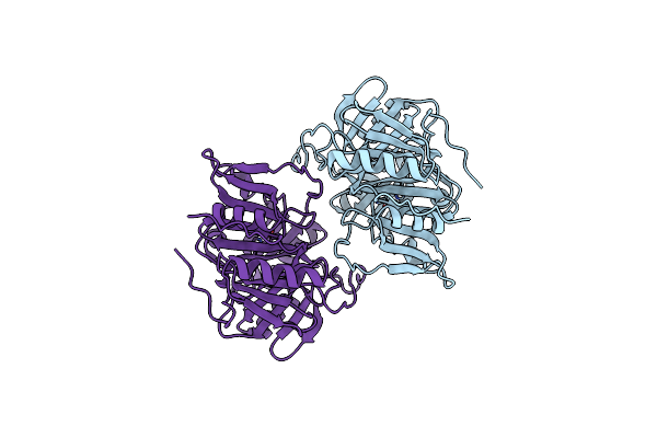 |
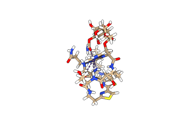
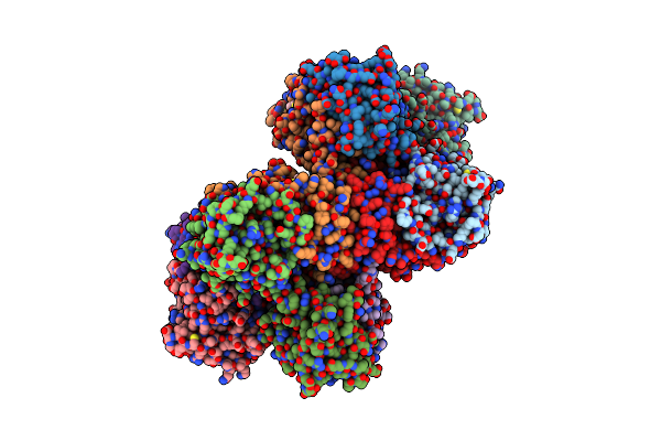
Crystal Structure Of Homoprotocatechuate 2,3-Dioxygenase From Arthrobacter Globiformis (Native, Non-Cryo)
Organism: Arthrobacter globiformis
Method: X-RAY DIFFRACTION Resolution:1.80 Å Release Date: 2003-06-10 Classification: OXIDOREDUCTASE Ligands: MN |
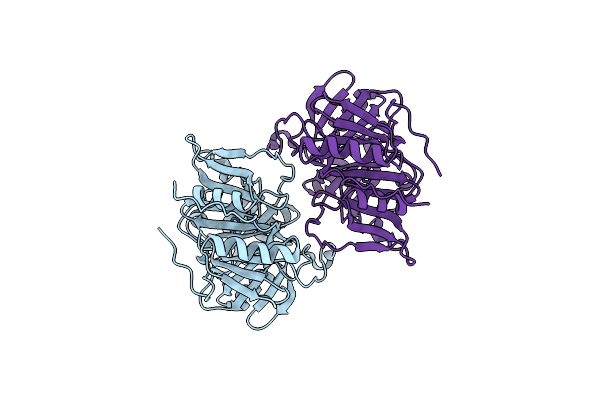 |
Crystal Structure Of Homoprotocatechuate 2,3-Dioxygenase From Arthrobacter Globiformis (Native, Low Temperature)
Organism: Arthrobacter globiformis
Method: X-RAY DIFFRACTION Resolution:1.50 Å Release Date: 2003-06-10 Classification: OXIDOREDUCTASE Ligands: MN |
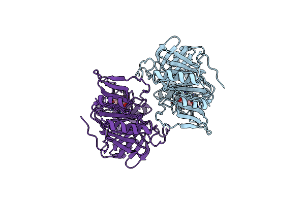 |
Anaerobic Substrate Complex Of Homoprotocatechuate 2,3-Dioxygenase From Arthrobacter Globiformis. (Complex With 3,4-Dihydroxyphenylacetate)
Organism: Arthrobacter globiformis
Method: X-RAY DIFFRACTION Resolution:1.90 Å Release Date: 2003-06-10 Classification: OXIDOREDUCTASE Ligands: MN, DHY |
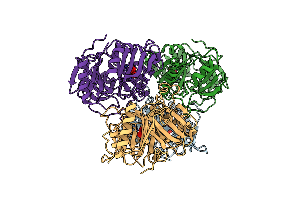 |
Crystal Structure Of Homoprotocatechuate 2,3-Dioxygenase From Brevibacterium Fuscum
Organism: Brevibacterium fuscum
Method: X-RAY DIFFRACTION Resolution:1.60 Å Release Date: 2003-06-10 Classification: OXIDOREDUCTASE Ligands: FEL |
 |
 |
Structure Of Protocatechuate 3,4-Dioxygenase Complexed With 3,4-Dihydroxyphenylacetate
Organism: Pseudomonas putida
Method: X-RAY DIFFRACTION Resolution:2.40 Å Release Date: 1998-02-25 Classification: DIOXYGENASE Ligands: FE, BME, DHY |