
Search Count: 15
 |
Organism: Bradyrhizobium diazoefficiens
Method: X-RAY DIFFRACTION Resolution:1.30 Å Release Date: 2015-09-23 Classification: CHAPERONE Ligands: SIN |
 |
Organism: Bradyrhizobium diazoefficiens
Method: X-RAY DIFFRACTION Resolution:1.40 Å Release Date: 2015-09-23 Classification: CHAPERONE Ligands: CU |
 |
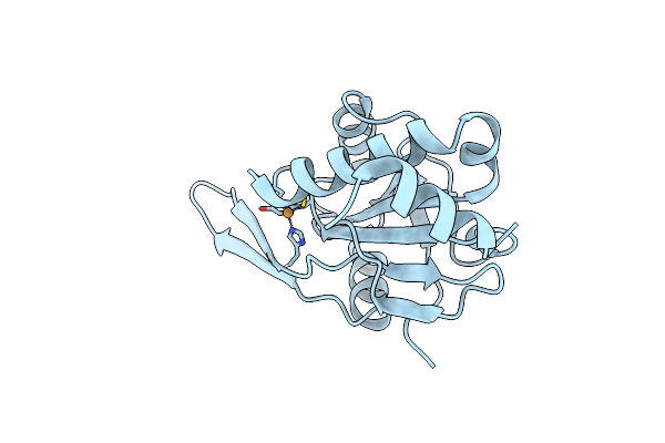
Crystal Structure Of The Periplasmic Domain Of Subunit Ii Of Cytochrome Oxidase (Coxb) Of Bradyrhizobium Japonicum
Organism: Bradyrhizobium diazoefficiens
Method: X-RAY DIFFRACTION Resolution:1.30 Å Release Date: 2015-09-09 Classification: OXIDOREDUCTASE Ligands: CU |
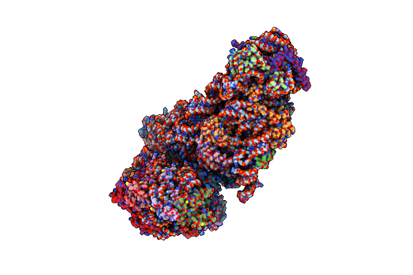 |
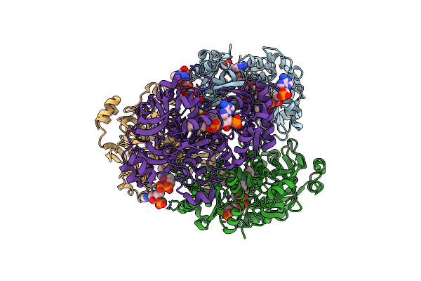
Organism: Hepatitis c virus, Homo sapiens
Method: ELECTRON MICROSCOPY Resolution:3.90 Å Release Date: 2015-07-15 Classification: RIBOSOME Ligands: MG, ZN |
 |
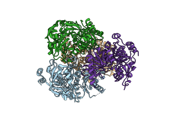
Organism: Candida tropicalis
Method: X-RAY DIFFRACTION Resolution:2.00 Å Release Date: 2015-03-18 Classification: OXIDOREDUCTASE Ligands: GOL, SO4 |
 |
Organism: Candida tropicalis
Method: X-RAY DIFFRACTION Resolution:1.70 Å Release Date: 2015-03-18 Classification: OXIDOREDUCTASE Ligands: NAP, COO |
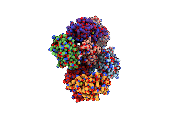 |
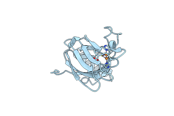
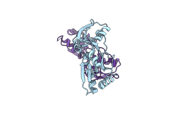
Crystal Structure Of The Mixed Disulfide Complex Of Thioredoxin-Like Tlpas(C110S) And Copper Chaperone Scois(C74S)
Organism: Bradyrhizobium diazoefficiens usda 110
Method: X-RAY DIFFRACTION Resolution:2.20 Å Release Date: 2014-10-01 Classification: OXIDODREDUCTASE/COPPER BINDING PROTEIN Ligands: SCN, PEG, NA |
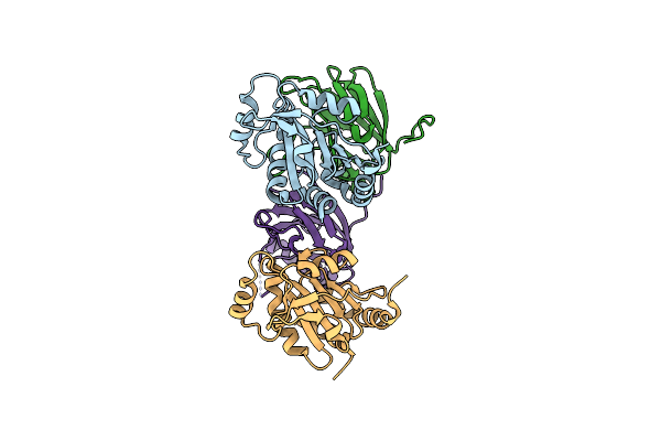 |
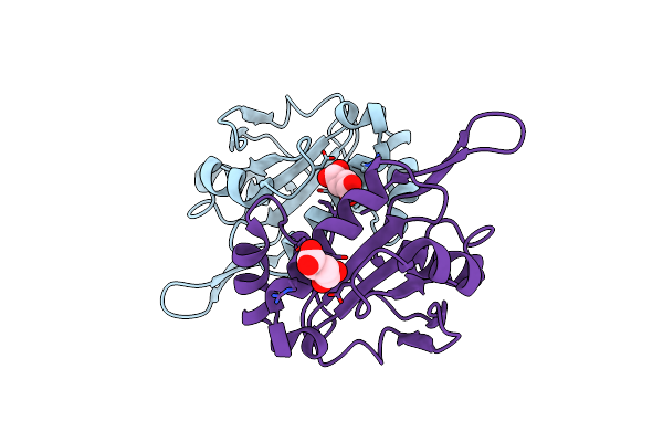
Crystal Structure Of The Mixed Disulfide Intermediate Between Thioredoxin-Like Tlpas(C110S) And Subunit Ii Of Cytochrome C Oxidase Coxbpd (C233S)
Organism: Bradyrhizobium diazoefficiens, Bradyrhizobium diazoefficiens (strain jcm 10833 / iam 13628 / nbrc 14792 / usda 110)
Method: X-RAY DIFFRACTION Resolution:2.00 Å Release Date: 2014-10-01 Classification: PROTEIN BINDING |
 |
Organism: Yersinia pseudotuberculosis
Method: X-RAY DIFFRACTION Resolution:2.40 Å Release Date: 2012-09-12 Classification: TRANSCRIPTION |
 |
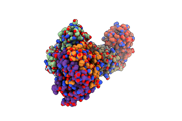
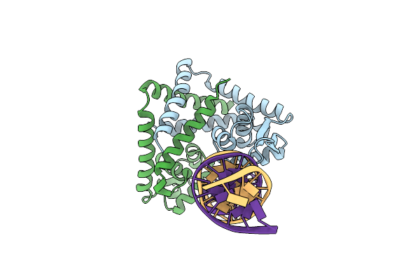
Crystal Structure Of Rova From Yersinia In Complex With A Rova Promoter Fragment
Organism: Yersinia pseudotuberculosis
Method: X-RAY DIFFRACTION Resolution:2.05 Å Release Date: 2012-09-12 Classification: TRANSCRIPTION |
 |
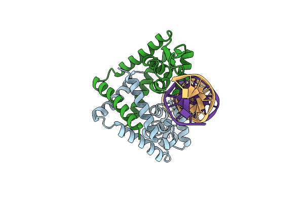
Crystal Structure Of Rova From Yersinia In Complex With An Invasin Promoter Fragment
Organism: Yersinia pseudotuberculosis
Method: X-RAY DIFFRACTION Resolution:1.85 Å Release Date: 2012-09-12 Classification: TRANSCRIPTION |
 |
Structure Of The 2-Octenoyl-Coa Carboxylase Reductase Cinf In Complex With Nadp And 2-Octenoyl-Coa
Organism: Streptomyces sp.
Method: X-RAY DIFFRACTION Resolution:1.90 Å Release Date: 2011-12-07 Classification: OXIDOREDUCTASE Ligands: CO8, NAP |
 |
Apo-Structure Of 2-Octenoyl-Coa Carboxylase Reductase Cinf From Streptomyces Sp.
Organism: Streptomyces sp.
Method: X-RAY DIFFRACTION Resolution:2.25 Å Release Date: 2011-12-07 Classification: OXIDOREDUCTASE |
 |
Effector Binding Domain Of Lysr-Type Transcription Factor Rovm From Y. Pseudotuberculosis
Organism: Yersinia pseudotuberculosis
Method: X-RAY DIFFRACTION Resolution:2.40 Å Release Date: 2011-01-26 Classification: TRANSCRIPTION |
 |
Crystal Structure Of Mexz, A Key Repressor Responsible For Antibiotic Resistance In Pseudomonas Aeruginosa.
Organism: Pseudomonas aeruginosa
Method: X-RAY DIFFRACTION Resolution:2.90 Å Release Date: 2010-08-18 Classification: TRANSCRIPTION |