
Search Count: 30
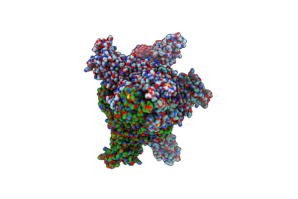 |
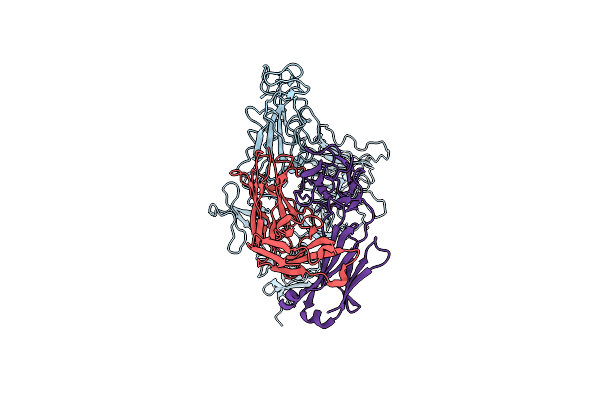
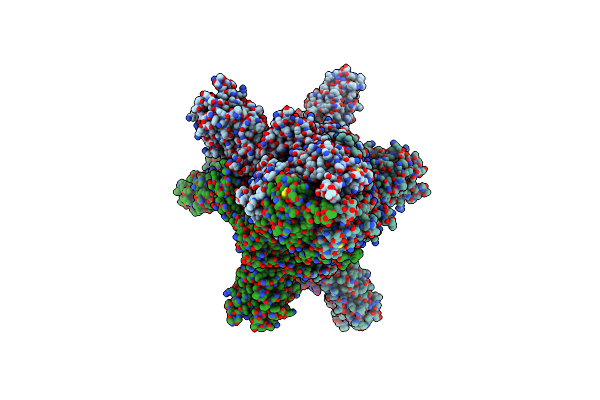
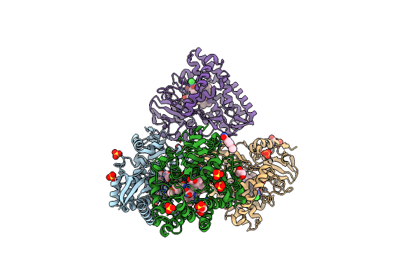
State 2 Of Sars-Cov-2 Xbb Variant Spike Protein Trimer Complexed With Antibody Pw5-5
Organism: Severe acute respiratory syndrome coronavirus 2, Homo sapiens
Method: ELECTRON MICROSCOPY Release Date: 2024-09-04 Classification: VIRAL PROTEIN/IMMUNE SYSTEM |
 |
Organism: Severe acute respiratory syndrome coronavirus 2, Homo sapiens
Method: ELECTRON MICROSCOPY Release Date: 2024-08-14 Classification: VIRAL PROTEIN/IMMUNE SYSTEM |
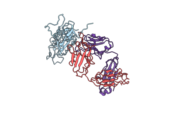 |
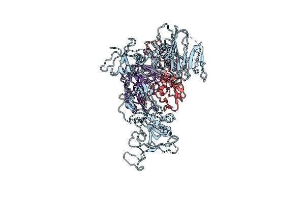
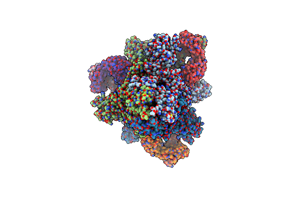
The Local Refined Map Of Sars-Cov-2 Xbb Variant Spike Protein Complexed With Antibody Pw5-535
Organism: Homo sapiens, Severe acute respiratory syndrome coronavirus 2
Method: ELECTRON MICROSCOPY Release Date: 2024-08-14 Classification: IMMUNE SYSTEM/VIRAL PROTEIN |
 |
Organism: Homo sapiens, Severe acute respiratory syndrome coronavirus 2
Method: ELECTRON MICROSCOPY Release Date: 2024-08-14 Classification: IMMUNE SYSTEM/VIRAL PROTEIN |
 |
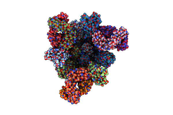
The Local Refined Map Of Sars-Cov Spike Protein Complexed With Antibody Pw5-5
Organism: Severe acute respiratory syndrome coronavirus 2, Homo sapiens
Method: ELECTRON MICROSCOPY Release Date: 2024-08-14 Classification: VIRAL PROTEIN/IMMUNE SYSTEM |
 |
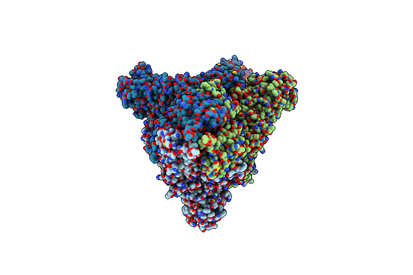
Monomer State Of Sars-Cov-2 Xbb Variant Spike Protein Trimer Complexed With Antibody Pw5-5
Organism: Homo sapiens, Severe acute respiratory syndrome coronavirus 2
Method: ELECTRON MICROSCOPY Release Date: 2024-08-14 Classification: IMMUNE SYSTEM/VIRAL PROTEIN |
 |
Organism: Homo sapiens, Severe acute respiratory syndrome coronavirus 2
Method: ELECTRON MICROSCOPY Release Date: 2024-08-14 Classification: IMMUNE SYSTEM/VIRAL PROTEIN |
 |
Organism: Severe acute respiratory syndrome coronavirus 2, Homo sapiens
Method: ELECTRON MICROSCOPY Release Date: 2024-08-14 Classification: VIRAL PROTEIN/IMMUNE SYSTEM |
 |
The Local Refined Map Of Sars-Cov-2 Omicron Ba.1 Spike Complexed With Antibody Pw5-570
Organism: Homo sapiens, Severe acute respiratory syndrome coronavirus 2
Method: ELECTRON MICROSCOPY Release Date: 2024-08-14 Classification: IMMUNE SYSTEM/VIRAL PROTEIN |
 |
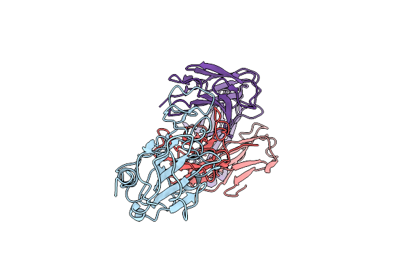
State 1 Of Sars-Cov-2 Xbb Variant Spike Protein Trimer Complexed With Antibody Pw5-5
Organism: Severe acute respiratory syndrome coronavirus 2, Homo sapiens
Method: ELECTRON MICROSCOPY Release Date: 2024-08-14 Classification: VIRAL PROTEIN/IMMUNE SYSTEM |
 |
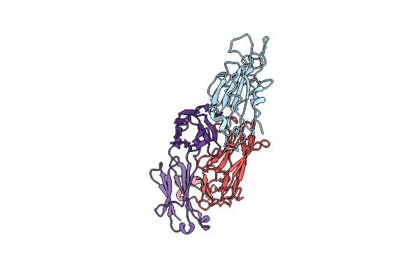
Structure Of Sars-Cov-2 Xbb Variant Spike Protein Complexed With Broadly Neutralizing Antibody Pw5-535
Organism: Severe acute respiratory syndrome coronavirus 2, Homo sapiens
Method: ELECTRON MICROSCOPY Release Date: 2024-08-14 Classification: IMMUNE SYSTEM/VIRAL PROTEIN |
 |
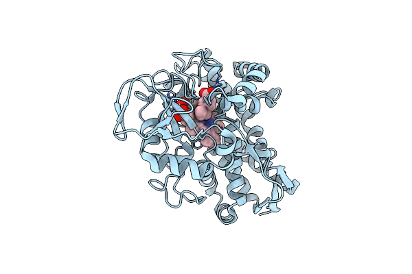
Organism: Rhodopseudomonas palustris haa2
Method: X-RAY DIFFRACTION Resolution:2.30 Å Release Date: 2023-08-30 Classification: OXIDOREDUCTASE Ligands: MH0, HX1, CL |
 |
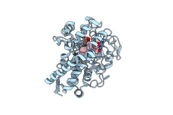
Organism: Rhodopseudomonas palustris haa2
Method: X-RAY DIFFRACTION Resolution:1.54 Å Release Date: 2023-01-04 Classification: OXIDOREDUCTASE Ligands: HEM, 8ZU, CL |
 |
Organism: Rhodopseudomonas palustris (strain haa2)
Method: X-RAY DIFFRACTION Resolution:1.37 Å Release Date: 2022-06-29 Classification: OXIDOREDUCTASE Ligands: HEM, 8ZU, CL |
 |
Organism: Rhodopseudomonas palustris (strain haa2)
Method: X-RAY DIFFRACTION Resolution:1.38 Å Release Date: 2019-02-27 Classification: OXIDOREDUCTASE Ligands: HEM, 8ZU, CL |
 |
Organism: Rhodopseudomonas palustris (strain haa2)
Method: X-RAY DIFFRACTION Resolution:2.30 Å Release Date: 2019-02-27 Classification: OXIDOREDUCTASE Ligands: HEM, 8ZX, CL, GOL, SO4 |
 |
Model Of Human Anaphase-Promoting Complex/Cyclosome (Apc/C-Cdh1) With E2 Ube2S Poised For Polyubiquitination Where Ube2S, Apc2, And Apc11 Are Modeled Into Low Resolution Density
Organism: Homo sapiens, Saccharomyces cerevisiae (strain atcc 204508 / s288c)
Method: ELECTRON MICROSCOPY Release Date: 2016-10-26 Classification: CELL CYCLE |
 |
Model Of Human Anaphase-Promoting Complex/Cyclosome (Apc/C-Cdh1) With A Cross Linked Ubiquitin Variant-Substrate-Ube2C (Ubch10) Complex Representing Key Features Of Multiubiquitination
Organism: Homo sapiens, Saccharomyces cerevisiae, Rattus norvegicus
Method: ELECTRON MICROSCOPY Release Date: 2016-09-14 Classification: SIGNALING PROTEIN |
 |
Model Of Human Anaphase-Promoting Complex/Cyclosome (Apc15 Deletion Mutant), In Complex With The Mitotic Checkpoint Complex (Apc/C-Cdc20-Mcc) Based On Cryo Em Data At 4.8 Angstrom Resolution
Organism: Homo sapiens
Method: ELECTRON MICROSCOPY Release Date: 2016-09-07 Classification: CELL CYCLE |
 |
Model Of Human Anaphase-Promoting Complex/Cyclosome Complex (Apc15 Deletion Mutant) In Complex With The E2 Ube2C/Ubch10 Poised For Ubiquitin Ligation To Substrate (Apc/C-Cdc20-Substrate-Ube2C)
Organism: Homo sapiens, Saccharomyces cerevisiae
Method: ELECTRON MICROSCOPY Release Date: 2016-08-24 Classification: CELL CYCLE |