
Search Count: 33
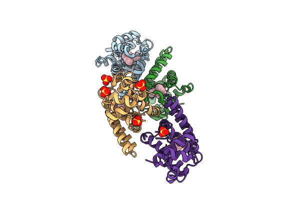 |
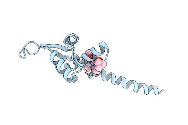
Organism: Salmonella enterica subsp. enterica serovar typhimurium str. 14028s
Method: X-RAY DIFFRACTION Release Date: 2025-10-22 Classification: PHOTOSYNTHESIS Ligands: A1EKZ, SO4 |
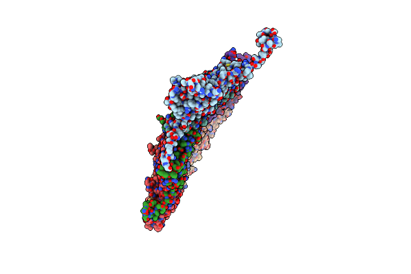 |
Organism: Xanthomonas phage phixacjx1
Method: ELECTRON MICROSCOPY Release Date: 2025-05-07 Classification: VIRUS |
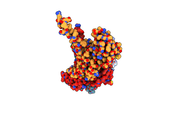 |
The Composite Cryo-Em Structure Of The Head-To-Tail Connector And Head-Proximal Tail Components Of Bacteriophage Phixacjx1
Organism: Xanthomonas phage phixacjx1
Method: ELECTRON MICROSCOPY Release Date: 2025-05-07 Classification: VIRUS |
 |
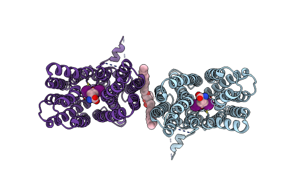
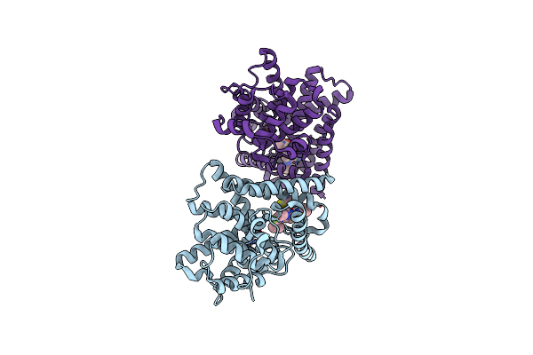
Cryoem Structure Of Thyroid Hormone Transporter Mct8 Bound With Silychristin
Organism: Homo sapiens
Method: ELECTRON MICROSCOPY Release Date: 2025-04-02 Classification: MEMBRANE PROTEIN Ligands: A1IET, PC1, DU0 |
 |
Organism: Homo sapiens
Method: ELECTRON MICROSCOPY Release Date: 2025-04-02 Classification: MEMBRANE PROTEIN Ligands: T3, PC1 |
 |
Crystal Structure Of Human Phosphodiesterase 10A In Complex With N-(2-Amino-2-Thioxoethyl)-2-(3-(3-(Dimethylcarbamoyl)-6-Fluoroimidazo[1,2-A]Pyridin-2-Yl)Azetidin-1-Yl)Quinoline-4-Carboxamide
Organism: Homo sapiens
Method: X-RAY DIFFRACTION Resolution:2.40 Å Release Date: 2025-01-29 Classification: HYDROLASE Ligands: A1L31, ZN, MG |
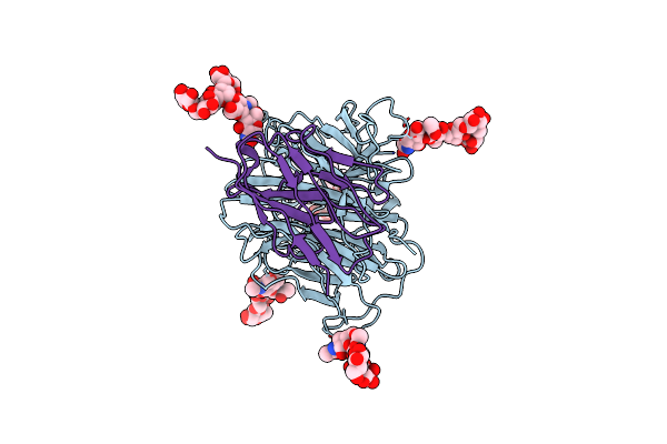 |
Structure Of Nipah Virus Bangladesh String G Protein Ectodomain Monomer Bound To Single-Domain Antibody N425 At 3.22 Angstroms Overall Resolution
Organism: Homo sapiens, Henipavirus nipahense
Method: ELECTRON MICROSCOPY Release Date: 2024-09-18 Classification: VIRAL PROTEIN |
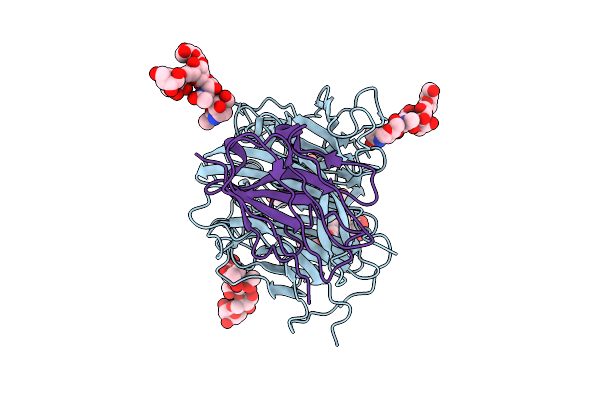 |
Structure Of Nipah Virus Malaysia String G Protein Ectodomain Monomer Bound To Single-Domain Antibody N425 At 3.63 Angstroms Overall Resolution
Organism: Henipavirus nipahense, Homo sapiens
Method: ELECTRON MICROSCOPY Release Date: 2024-09-18 Classification: VIRAL PROTEIN |
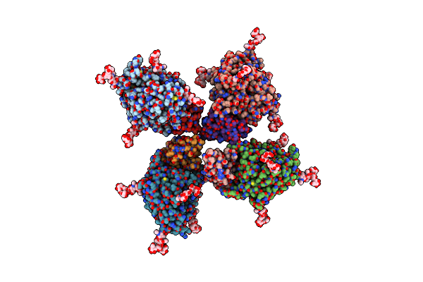 |
Structure Of Nipah Virus Bangladesh String G Protein Ectodomain Tetramer Bound To Single-Domain Antibody N425 At 5.87 Angstroms Overall Resolution
Organism: Henipavirus nipahense, Homo sapiens
Method: ELECTRON MICROSCOPY Release Date: 2024-09-18 Classification: VIRAL PROTEIN |
 |
Organism: Severe acute respiratory syndrome coronavirus 2, Synthetic construct
Method: ELECTRON MICROSCOPY Release Date: 2024-07-24 Classification: VIRAL PROTEIN/IMMUNE SYSTEM |
 |
Organism: Lactococcus lactis subsp. cremoris (strain mg1363)
Method: X-RAY DIFFRACTION Resolution:2.60 Å Release Date: 2024-07-17 Classification: PHOTOSYNTHESIS Ligands: U1O |
 |
Organism: Chryseobacterium
Method: ELECTRON MICROSCOPY Release Date: 2024-06-05 Classification: DNA BINDING PROTEIN/DNA/RNA |
 |
Organism: Chryseobacterium
Method: ELECTRON MICROSCOPY Release Date: 2024-06-05 Classification: DNA BINDING PROTEIN/DNA/RNA |
 |
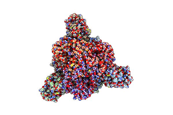
Structure Of Cbcas9-Pcriic1 Complex Bound To 28-Bp Dna Substrate (20-Nt Complementary)
Organism: Chryseobacterium
Method: ELECTRON MICROSCOPY Release Date: 2024-06-05 Classification: DNA BINDING PROTEIN/DNA/RNA Ligands: MG |
 |
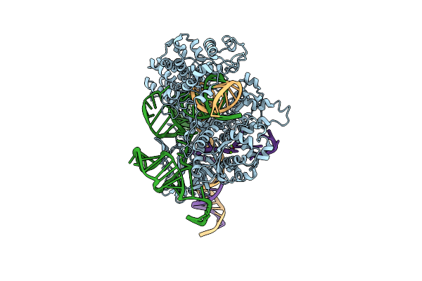
Structure Of Cbcas9-Pcriic1 Complex Bound To 62-Bp Dna Substrate (Symmetric 20-Nt Complementary)
Organism: Chryseobacterium
Method: ELECTRON MICROSCOPY Release Date: 2024-06-05 Classification: DNA BINDING PROTEIN/DNA/RNA Ligands: MG |
 |
Structure Of Cbcas9-Pcriic1 Complex Bound To 62-Bp Dna Substrate (Non-Targeting Complex)
Organism: Chryseobacterium
Method: ELECTRON MICROSCOPY Release Date: 2024-06-05 Classification: DNA BINDING PROTEIN/DNA/RNA Ligands: MG |
 |
Organism: Staphylococcus aureus, Synthetic construct
Method: ELECTRON MICROSCOPY Release Date: 2023-03-15 Classification: DNA BINDING PROTEIN/RNA/DNA Ligands: MG |
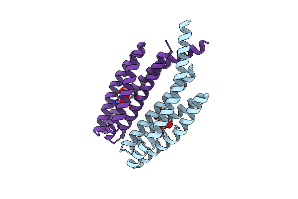 |
Organism: Comamonas testosteroni cnb-2
Method: X-RAY DIFFRACTION Resolution:1.80 Å Release Date: 2023-01-04 Classification: SIGNALING PROTEIN Ligands: LMR |
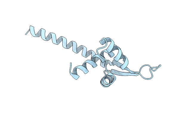 |
Organism: Lactococcus lactis subsp. cremoris mg1363
Method: X-RAY DIFFRACTION Resolution:2.60 Å Release Date: 2022-09-28 Classification: PHOTOSYNTHESIS |
 |
Organism: Lactococcus lactis subsp. cremoris mg1363
Method: X-RAY DIFFRACTION Resolution:2.50 Å Release Date: 2022-09-28 Classification: PHOTOSYNTHESIS Ligands: I1A |