
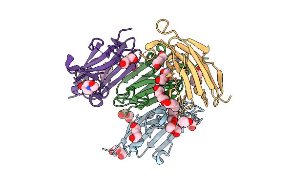
Search Count: 33
 |
Organism: Xenopus laevis, Homo sapiens, Synthetic construct
Method: ELECTRON MICROSCOPY Release Date: 2025-08-06 Classification: IMMUNE SYSTEM Ligands: NAG |
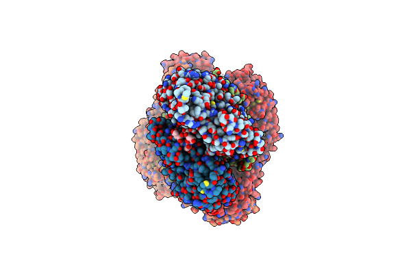
 |
Organism: Xenopus laevis, Synthetic construct
Method: ELECTRON MICROSCOPY Release Date: 2025-08-06 Classification: NUCLEAR PROTEIN |
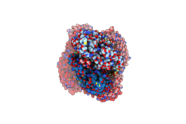
 |
Organism: Xenopus laevis, Synthetic construct, Homo sapiens
Method: ELECTRON MICROSCOPY Release Date: 2025-08-06 Classification: IMMUNE SYSTEM Ligands: NAG, HEM |
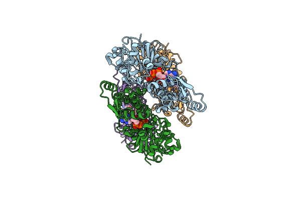
 |
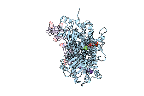
Native Dimeric Myeloperoxidase Bound To Nucleosome Core Particle; Composite Map
Organism: Xenopus laevis, Synthetic construct, Homo sapiens
Method: ELECTRON MICROSCOPY Release Date: 2025-08-06 Classification: IMMUNE SYSTEM Ligands: CL, HEM, NAG, CA |
 |
Native Dimeric Myeloperoxidase Bound To Nucleosome Core Particle, Intermediate State; Composite Map
Organism: Xenopus laevis, Synthetic construct, Homo sapiens
Method: ELECTRON MICROSCOPY Release Date: 2025-08-06 Classification: IMMUNE SYSTEM Ligands: CL, HEM, NAG, CA |
 |
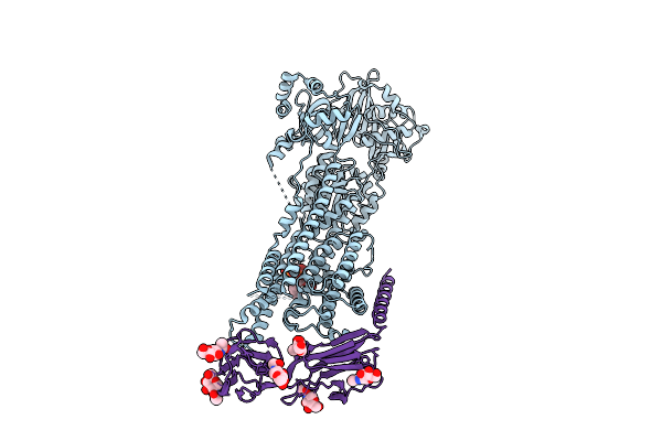
Organism: Mus musculus
Method: ELECTRON MICROSCOPY Release Date: 2025-08-06 Classification: MEMBRANE PROTEIN Ligands: MG, KXP, BEF, NAG |
 |
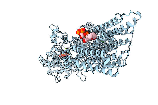
Cryo-Em Structure Of Mouse Pmca-Nptn Complex Captured In E1 State Without Calcium
Organism: Mus musculus
Method: ELECTRON MICROSCOPY Release Date: 2025-08-06 Classification: MEMBRANE PROTEIN Ligands: KXP, NAG |
 |
Organism: Mus musculus
Method: ELECTRON MICROSCOPY Release Date: 2025-08-06 Classification: MEMBRANE PROTEIN Ligands: ANP, MG, KXP, NAG |
 |
Cryo-Em Structure Of Mouse Pmca-Nptn Complex Captured In E2-Pi State (Alf4)
Organism: Mus musculus
Method: ELECTRON MICROSCOPY Release Date: 2025-08-06 Classification: MEMBRANE PROTEIN Ligands: ALF, MG, NAG |
 |
Organism: Mus musculus
Method: ELECTRON MICROSCOPY Release Date: 2025-08-06 Classification: MEMBRANE PROTEIN Ligands: ANP, CA, MG, KXP, NAG |
 |
Cryo-Em Structure Of Mouse Pmca Captured In E1-Atp In The Presence Of Calcium
Organism: Mus musculus
Method: ELECTRON MICROSCOPY Release Date: 2025-08-06 Classification: MEMBRANE PROTEIN Ligands: ANP, CA, MG, KXP |
 |
Organism: Mus musculus
Method: ELECTRON MICROSCOPY Release Date: 2025-08-06 Classification: MEMBRANE PROTEIN Ligands: BEF, MG, KXP |
 |
Organism: Mus musculus
Method: ELECTRON MICROSCOPY Release Date: 2025-08-06 Classification: MEMBRANE PROTEIN Ligands: CA, KXP, NAG |
 |
Organism: Mus musculus
Method: X-RAY DIFFRACTION Release Date: 2025-08-06 Classification: CELL ADHESION Ligands: PEG, EPE |
 |
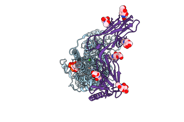
Nucleosome Core Particle Bound By One Molecule Of Dtt-Reduced Native Monomeric Myeloperoxidase
Organism: Xenopus laevis, Synthetic construct, Homo sapiens
Method: ELECTRON MICROSCOPY Release Date: 2025-08-06 Classification: IMMUNE SYSTEM Ligands: HEM, NAG |
 |
Nucleosome Core Particle Bound By Two Molecules Of Dtt-Reduced Native Monomeric Myeloperoxidase
Organism: Xenopus laevis, Synthetic construct, Homo sapiens
Method: ELECTRON MICROSCOPY Release Date: 2025-08-06 Classification: IMMUNE SYSTEM Ligands: HEM, NAG |
 |
Nucleosome Core Particle Bound By One Monomer And One Dimer Of Of Dtt-Reduced Native Myeloperoxidase
Organism: Xenopus laevis, Synthetic construct, Homo sapiens
Method: ELECTRON MICROSCOPY Release Date: 2025-08-06 Classification: IMMUNE SYSTEM Ligands: HEM, NAG |
 |
Structure Of The Formin Cdc12 Bound To The Barbed End Of Phalloidin-Stabilized F-Actin.
Organism: Homo sapiens, Schizosaccharomyces pombe, Amanita phalloides
Method: ELECTRON MICROSCOPY Release Date: 2024-04-10 Classification: STRUCTURAL PROTEIN Ligands: ADP, MG, PO4 |
 |
Structure Of The F-Actin Barbed End Bound By Cdc12 And Profilin (Ring Complex) At A Resolution Of 6.3 Angstrom
Organism: Homo sapiens, Schizosaccharomyces pombe, Amanita phalloides
Method: ELECTRON MICROSCOPY Release Date: 2024-04-10 Classification: STRUCTURAL PROTEIN Ligands: ADP, MG, PO4 |
 |
Organism: Oryctolagus cuniculus
Method: ELECTRON MICROSCOPY Release Date: 2024-04-10 Classification: STRUCTURAL PROTEIN Ligands: ADP, MG, PO4, ATP |