
Search Count: 21
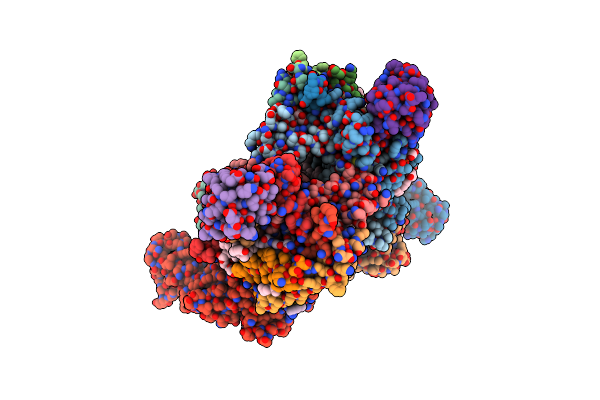 |
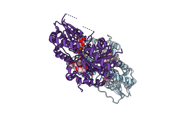
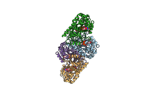
2.8 Angstrom Cryo-Em Structure Of The Dimeric Cytochrome B6F-Petp Complex From Synechocystis Sp. Pcc 6803 With Natively Bound Lipids And Plastoquinone Molecules
Organism: Synechocystis, Synechocystis sp. pcc 6803
Method: ELECTRON MICROSCOPY Release Date: 2022-07-06 Classification: PHOTOSYNTHESIS Ligands: HEM, PGV, ECH, PL9, CLA, HEC, LMG, 6PL, 2WA, FES, SQD, LFA |
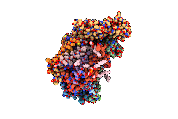 |
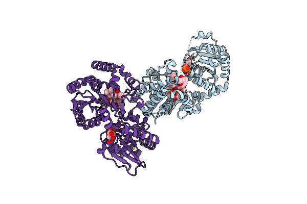
3.15 Angstrom Cryo-Em Structure Of The Dimeric Cytochrome B6F Complex From Synechocystis Sp. Pcc 6803 With Natively Bound Plastoquinone And Lipid Molecules.
Organism: Synechocystis sp. pcc 6803
Method: ELECTRON MICROSCOPY Release Date: 2022-07-06 Classification: OXIDOREDUCTASE Ligands: ECH, HEM, HEC, CLA, PGV, LFA, FES |
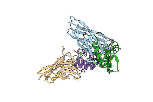 |
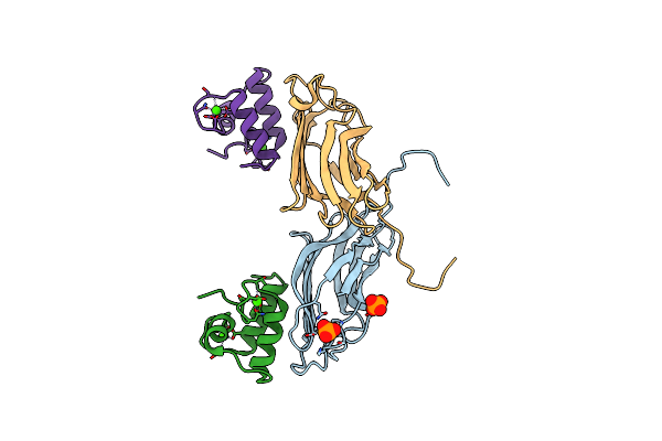
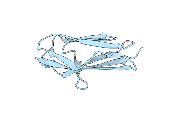
The Clostridium Cellulolyticum Dockerin Displays A Dual Binding Mode For Its Cohesin Partner
Organism: Clostridium cellulolyticum
Method: X-RAY DIFFRACTION Resolution:1.90 Å Release Date: 2008-05-20 Classification: CELL ADHESION Ligands: CA |
 |
The Clostridium Cellulolyticum Dockerin Displays A Dual Binding Mode For Its Cohesin Partner
Organism: Clostridium cellulolyticum
Method: X-RAY DIFFRACTION Resolution:1.49 Å Release Date: 2008-05-13 Classification: CELL ADHESION Ligands: CA |
 |
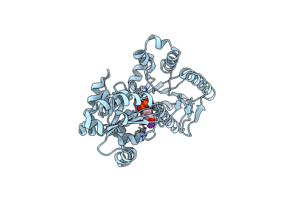
The Crystal Structure Of Macrolide Glycosyltransferases: A Blueprint For Antibiotic Engineering
Organism: Streptomyces antibioticus
Method: X-RAY DIFFRACTION Resolution:1.70 Å Release Date: 2007-03-27 Classification: TRANSFERASE Ligands: UDP, ZIO |
 |
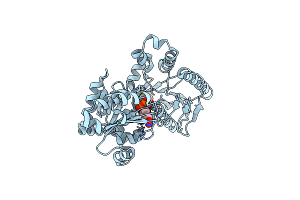
The Crystal Structure Of Macrolide Glycosyltransferases: A Blueprint For Antibiotic Engineering
Organism: Streptomyces antibioticus
Method: X-RAY DIFFRACTION Resolution:1.70 Å Release Date: 2007-03-27 Classification: TRANSFERASE Ligands: ERY, UDP, MG |
 |
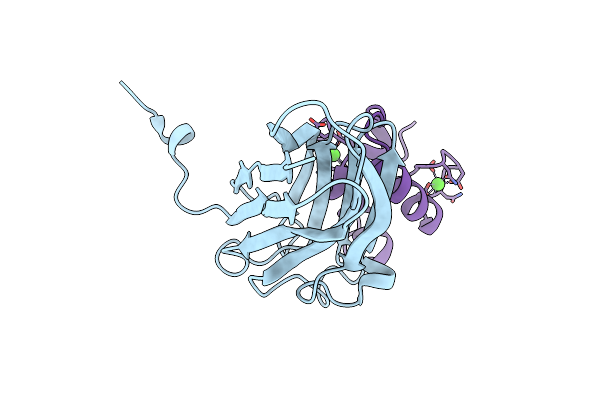
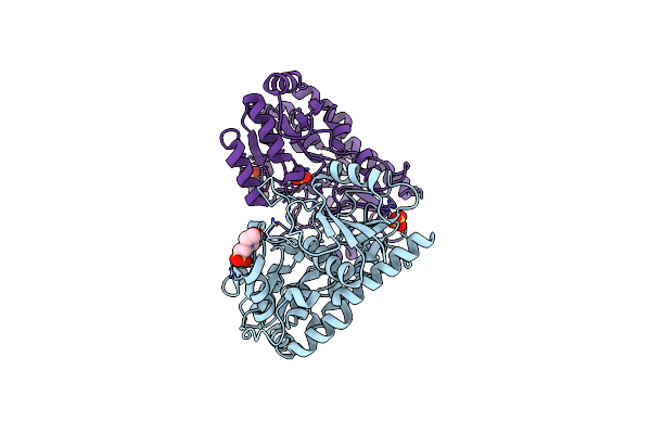
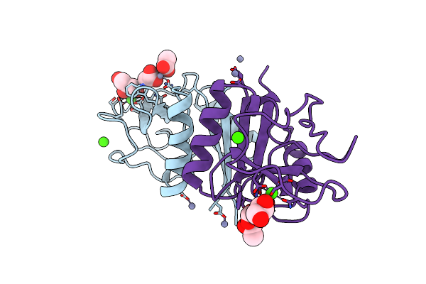
The S45A, T46A Mutant Of The Type I Cohesin-Dockerin Complex From The Cellulosome Of Clostridium Thermocellum
Organism: Clostridium thermocellum
Method: X-RAY DIFFRACTION Resolution:2.03 Å Release Date: 2007-02-13 Classification: CELL ADHESION Ligands: PO4, CA |
 |
Crystal Structure Of Avigt4, A Glycosyltransferase Involved In Avilamycin A Biosynthesis
Organism: Streptomyces viridochromogenes
Method: X-RAY DIFFRACTION Resolution:2.10 Å Release Date: 2006-10-11 Classification: TRANSFERASE Ligands: MES, SO4 |
 |
Crystal Structure Of Avigt4, A Glycosyltransferase Involved In Avilamycin A Biosynthesis
Organism: Streptomyces viridochromogenes
Method: X-RAY DIFFRACTION Resolution:2.30 Å Release Date: 2006-10-11 Classification: TRANSFERASE Ligands: UDP, GOL |
 |
Crystal Structure Of Waag, A Glycosyltransferase Involved In Lipopolysaccharide Biosynthesis
Organism: Escherichia coli
Method: X-RAY DIFFRACTION Resolution:1.60 Å Release Date: 2006-10-11 Classification: TRANSFERASE Ligands: UDP |
 |
Crystal Structure Of Waag, A Glycosyltransferase Involved In Lipopolysaccharide Biosynthesis
Organism: Escherichia coli
Method: X-RAY DIFFRACTION Resolution:1.50 Å Release Date: 2006-10-11 Classification: TRANSFERASE Ligands: U2F |
 |
Organism: Bacillus subtilis
Method: X-RAY DIFFRACTION Resolution:1.50 Å Release Date: 2005-02-16 Classification: HYDROLASE Ligands: CA, EDO |
 |
 |
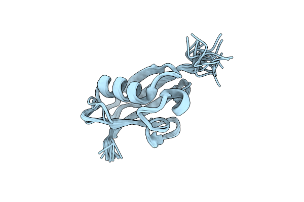
Nmr Structure Of The Complex Between The Third Dsrbd From Drosophila Staufen And A Rna Hairpin
Organism: Drosophila melanogaster
Method: SOLUTION NMR Release Date: 2000-08-21 Classification: cell cycle/RNA |
 |
Structure Of Tc14; A C-Type Lectin From The Tunicate Polyandrocarpa Misakiensis
Organism: Polyandrocarpa misakiensis
Method: X-RAY DIFFRACTION Resolution:2.00 Å Release Date: 1999-07-23 Classification: SUGAR BINDING PROTEIN Ligands: CA, ZN, ACT, GOL |
 |
Organism: Polyandrocarpa misakiensis
Method: X-RAY DIFFRACTION Resolution:2.20 Å Release Date: 1999-07-23 Classification: LECTIN Ligands: GAL, CA, ZN |
 |
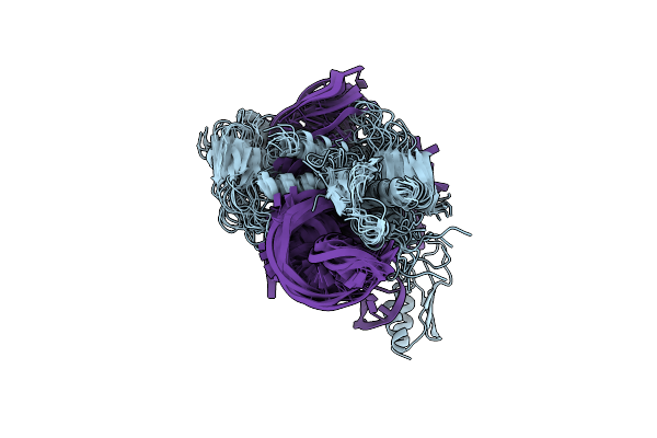
Organism: Thermus thermophilus
Method: X-RAY DIFFRACTION Resolution:1.95 Å Release Date: 1998-11-30 Classification: RIBOSOME |
 |
Organism: Escherichia coli
Method: SOLUTION NMR Release Date: 1997-04-01 Classification: S1 RNA-BINDING DOMAIN |
 |
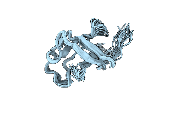
Twitchin Immunoglobulin Superfamily Domain (Igsf Module) (Ig 18'), Nmr, Minimized Average Structure
Organism: Caenorhabditis elegans
Method: SOLUTION NMR Release Date: 1996-12-23 Classification: MUSCLE PROTEIN |
 |
Twitchin Immunoglobulin Superfamily Domain (Igsf Module) (Ig 18'), Nmr, 30 Structures
Organism: Caenorhabditis elegans
Method: SOLUTION NMR Release Date: 1996-12-23 Classification: MUSCLE PROTEIN |