
Search Count: 38
 |
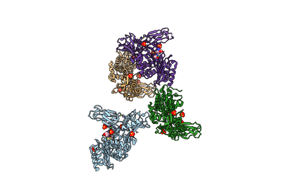
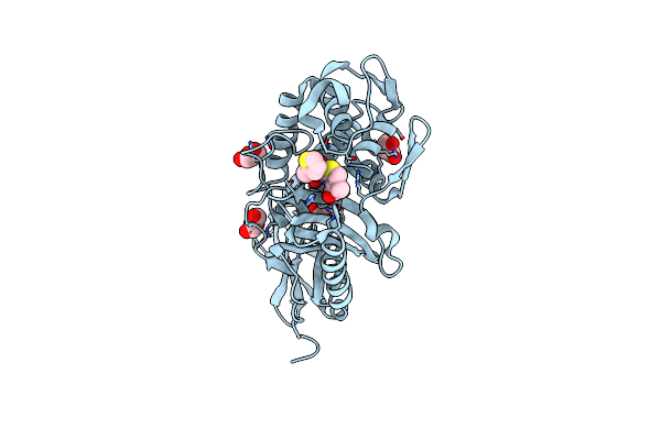
Crystal Structure Of The New Delhi Metallo-Beta-Lactamase-1 Adduct With A Lysine-Targeted Affinity Label
Organism: Escherichia coli
Method: X-RAY DIFFRACTION Resolution:2.02 Å Release Date: 2019-06-12 Classification: HYDROLASE Ligands: ZN, CA, N9M, N9J |
 |
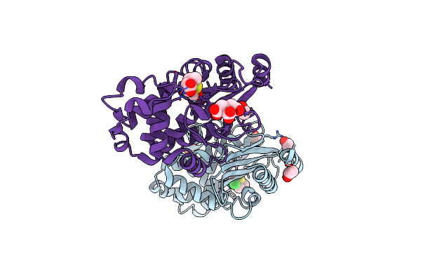
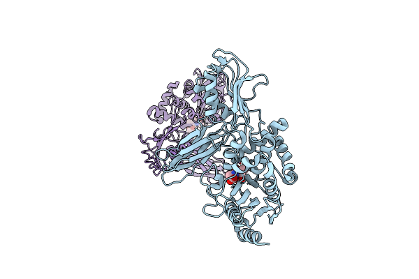
Crystal Structure Of A Complex Between Actinomadura R39 Dd-Peptidase And A Trifluoroketone Inhibitor
Organism: Actinomadura sp. r39
Method: X-RAY DIFFRACTION Resolution:2.60 Å Release Date: 2013-03-27 Classification: HYDROLASE Ligands: TFR, SO4, MG |
 |
Organism: Actinomadura sp. r39
Method: X-RAY DIFFRACTION Resolution:2.15 Å Release Date: 2013-03-20 Classification: HYDROLASE Ligands: IM2, MG, SO4, GOL, MES |
 |
Unexpected Tricovalent Binding Mode Of Boronic Acids Within The Active Site Of A Penicillin Binding Protein
Organism: Actinomadura sp.r39
Method: X-RAY DIFFRACTION Resolution:3.10 Å Release Date: 2012-02-29 Classification: HYDROLASE Ligands: B07, SO4, MG |
 |
Unexpected Tricovalent Binding Mode Of Boronic Acids Within The Active Site Of A Penicillin Binding Protein
Organism: Actinomadura sp. r39
Method: X-RAY DIFFRACTION Release Date: 2012-02-29 Classification: HYDROLASE Ligands: SO4, MG, FP5, ACN, 33D, MES |
 |
Unexpected Tricovalent Binding Mode Of Boronic Acids Within The Active Site Of A Penicillin Binding Protein
Organism: Actinomadura sp
Method: X-RAY DIFFRACTION Resolution:2.70 Å Release Date: 2011-07-27 Classification: HYDROLASE Ligands: BH6, SO4, MG |
 |
Unexpected Tricovalent Binding Mode Of Boronic Acids Within The Active Site Of A Penicillin Binding Protein
Organism: Actinomadura sp. r39
Method: X-RAY DIFFRACTION Release Date: 2011-07-27 Classification: HYDROLASE Ligands: FP5, SO4, MG, ACN |
 |
Unexpected Tricovalent Binding Mode Of Boronic Acids Within The Active Site Of A Penicillin Binding Protein
Organism: Actinomadura sp. r39
Method: X-RAY DIFFRACTION Resolution:2.50 Å Release Date: 2011-07-27 Classification: HYDROLASE Ligands: ZA3, SO4, MG |
 |
Structure Of Penicillin-Binding Protein 5 From E. Coli: Cloxacillin Acyl-Enzyme Complex
Organism: Escherichia coli
Method: X-RAY DIFFRACTION Resolution:1.90 Å Release Date: 2011-03-16 Classification: HYDROLASE/ANTIBIOTIC Ligands: CXV, GOL |
 |
Structure Of Penicillin-Binding Protein 5 From E.Coli: Cefoxitin Acyl-Enzyme Complex
Organism: Escherichia coli
Method: X-RAY DIFFRACTION Resolution:2.10 Å Release Date: 2011-03-16 Classification: HYDROLASE/ANTIBIOTIC Ligands: 1QL, GOL |
 |
Structure Of Penicillin-Binding Protein 5 From E. Coli: Imipenem Acyl-Enzyme Complex
Organism: Escherichia coli
Method: X-RAY DIFFRACTION Resolution:1.50 Å Release Date: 2011-03-16 Classification: HYDROLASE/ANTIBIOTIC Ligands: IM2, GOL |
 |
Crystal Structure Of A Complex Between Actinomadura R39 Dd Peptidase And A Peptidoglycan Mimetic Boronate Inhibitor
Organism: Actinomadura
Method: X-RAY DIFFRACTION Resolution:2.40 Å Release Date: 2010-07-21 Classification: HYDROLASE/INHIBITOR Ligands: SO4, CO, BO8, MES |
 |
Crystal Structure Of The Class A Beta-Lactamase Bs3 Inhibited By 6- Beta-Iodopenicillanate.
Organism: Bacillus licheniformis
Method: X-RAY DIFFRACTION Resolution:1.65 Å Release Date: 2009-12-01 Classification: HYDROLASE Ligands: BIY, CL, SO4, EOH, CIT |
 |
Crystal Structure Of The Actinomadura R39 Dd-Peptidase Inhibited By 6- Beta-Iodopenicillanate.
Organism: Actinomadura sp.
Method: X-RAY DIFFRACTION Resolution:2.20 Å Release Date: 2009-12-01 Classification: HYDROLASE Ligands: BIY, SO4, CO |
 |
Crystal Structure Of Actinomadura R39 Dd-Peptidase Complexed With A Peptidoglycan-Mimetic Cephalosporin
Organism: Actinomadura sp.
Method: X-RAY DIFFRACTION Resolution:2.40 Å Release Date: 2008-11-25 Classification: HYDROLASE Ligands: REC, SO4, MG |
 |
Crystal Structure Of Actinomadura R39 Dd-Peptidase Complexed With A Peptidoglycan-Mimetic Cephalosporin
Organism: Actinomadura sp.
Method: X-RAY DIFFRACTION Resolution:2.25 Å Release Date: 2008-11-25 Classification: HYDROLASE Ligands: REZ, SO4, MG |
 |
Crystal Structure Of E. Coli Penicillin-Binding Protein 5 In Complex With A Peptide-Mimetic Penicillin
Organism: Escherichia coli
Method: X-RAY DIFFRACTION Resolution:2.00 Å Release Date: 2008-08-26 Classification: HYDROLASE Ligands: HJ3, GOL |
 |
Crystal Structure Of E. Coli Penicillin-Binding Protein 5 In Complex With A Peptide-Mimetic Cephalosporin
Organism: Escherichia coli
Method: X-RAY DIFFRACTION Resolution:1.60 Å Release Date: 2008-08-26 Classification: HYDROLASE Ligands: GOL, HJ2 |
 |
Structure Of Ampc Beta-Lactamase With Cross-Linked Active Site After Exposure To Small Molecule Inhibitor
Organism: Escherichia coli
Method: X-RAY DIFFRACTION Resolution:1.80 Å Release Date: 2007-08-14 Classification: HYDROLASE Ligands: PO4 |
 |
Crystal Structure Of The Bacillus Subtilis Pbp4A, And Its Complex With A Peptidoglycan Mimetic Peptide.
Organism: Bacillus subtilis
Method: X-RAY DIFFRACTION Resolution:2.80 Å Release Date: 2007-07-03 Classification: HYDROLASE Ligands: REZ, DAL |