
Search Count: 18
 |
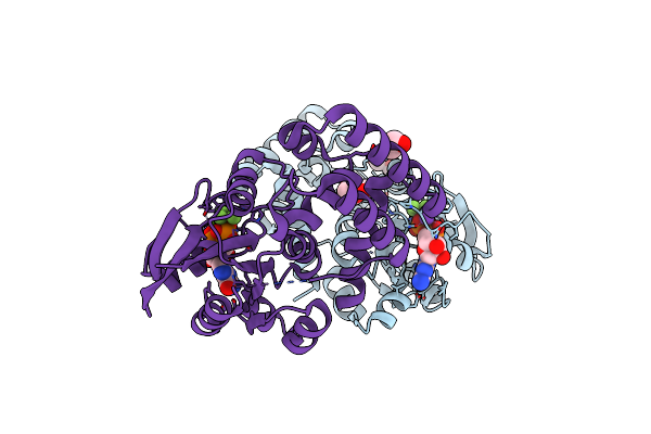
Organism: Aquifex aeolicus (strain vf5)
Method: X-RAY DIFFRACTION Resolution:2.18 Å Release Date: 2021-05-12 Classification: HYDROLASE Ligands: ALF, GDP, PEG, TRS, MG, K |
 |
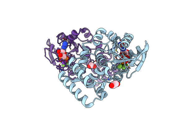
Organism: Streptococcus thermophilus (strain atcc baa-250 / lmg 18311)
Method: X-RAY DIFFRACTION Resolution:2.35 Å Release Date: 2021-04-21 Classification: HYDROLASE Ligands: GDP, ALF, MG, K, CL, PEG, EDO |
 |
Organism: Streptococcus thermophilus (strain atcc baa-250 / lmg 18311)
Method: X-RAY DIFFRACTION Resolution:2.50 Å Release Date: 2021-04-14 Classification: HYDROLASE Ligands: EDO, GDP, ALF, MG, K, CL |
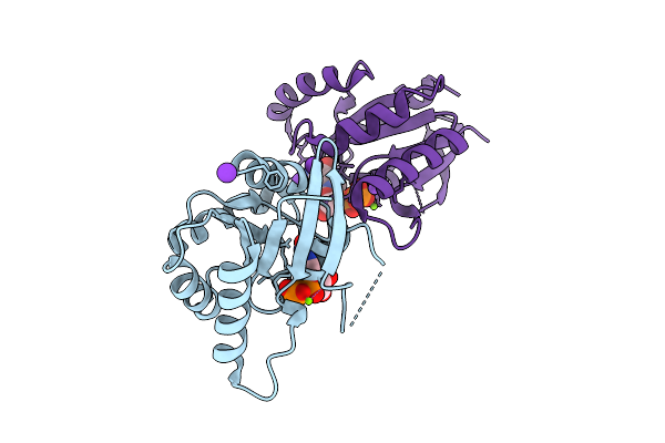 |
Organism: Bacillus subtilis (strain 168)
Method: X-RAY DIFFRACTION Resolution:1.50 Å Release Date: 2017-05-31 Classification: HYDROLASE Ligands: GDP, K, MG, NA |
 |
Organism: Mycobacterium tuberculosis
Method: X-RAY DIFFRACTION Resolution:1.98 Å Release Date: 2014-04-16 Classification: TRANSFERASE Ligands: ATP, MG, GN1, CO |
 |
Organism: Mycobacterium tuberculosis
Method: X-RAY DIFFRACTION Resolution:2.47 Å Release Date: 2013-08-07 Classification: TRANSFERASE Ligands: POP, MG, CO, UD1 |
 |
Organism: Mycobacterium tuberculosis
Method: X-RAY DIFFRACTION Resolution:1.90 Å Release Date: 2013-08-07 Classification: TRANSFERASE Ligands: POP, MG, CO, UD1 |
 |
Crystal Structure Of Glmu From Mycobacterium Tuberculosis In Complex With Uridine-Diphosphate-N-Acetylglucosamine And Pyrophosphate Snapshot 2
Organism: Mycobacterium tuberculosis
Method: X-RAY DIFFRACTION Resolution:2.04 Å Release Date: 2013-08-07 Classification: TRANSFERASE Ligands: UD1, GN1, POP, MG, CO |
 |
Organism: Mycobacterium tuberculosis
Method: X-RAY DIFFRACTION Resolution:2.03 Å Release Date: 2013-03-13 Classification: TRANSFERASE Ligands: MG, CO, UD1, POP |
 |
Crystal Structure Of Glmu From Mycobacterium Tuberculosis In Complex With Glucosamine-1-Phosphate
Organism: Mycobacterium tuberculosis
Method: X-RAY DIFFRACTION Resolution:2.60 Å Release Date: 2013-03-13 Classification: TRANSFERASE Ligands: GN1, CO, MG |
 |
Crystal Structure Of Glmu From Mycobacterium Tuberculosis In Complex With Acetyl Coenzyme A And Uridine-Diphosphate-N-Acetylglucosamine
Organism: Mycobacterium tuberculosis
Method: X-RAY DIFFRACTION Resolution:2.33 Å Release Date: 2012-08-08 Classification: TRANSFERASE Ligands: MG, ACO, UD1 |
 |
Crystal Structure Of Glmu From Mycobacterium Tuberculosis In Complex With Coenzyme A, Glucosamine 1-Phosphate And Uridine-Diphosphate-N-Acetylglucosamine
Organism: Mycobacterium tuberculosis
Method: X-RAY DIFFRACTION Resolution:1.98 Å Release Date: 2012-08-08 Classification: TRANSFERASE Ligands: MG, COA, GP1, UD1 |
 |
Crystal Structure Of N-Acetylglucosamine-1-Phosphate Uridyltransferase (Glmu) From Mycobacterium Tuberculosis In A Cubic Space Group.
Organism: Mycobacterium tuberculosis
Method: X-RAY DIFFRACTION Resolution:3.41 Å Release Date: 2009-06-09 Classification: TRANSFERASE Ligands: SO4 |
 |
Crystal Structure Of Glmu From Mycobacterium Tuberculosis In Complex With Uridine-Diphosphate-N-Acetylglucosamine.
Organism: Mycobacterium tuberculosis
Method: X-RAY DIFFRACTION Resolution:2.38 Å Release Date: 2009-05-19 Classification: TRANSFERASE Ligands: MG, CO, UD1 |
 |
Organism: Mycobacterium tuberculosis
Method: X-RAY DIFFRACTION Resolution:2.23 Å Release Date: 2009-05-19 Classification: TRANSFERASE Ligands: MG |
 |
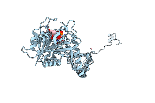
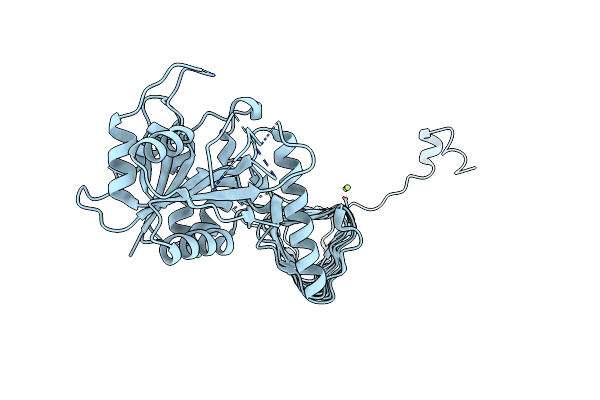
Organism: Mus musculus
Method: X-RAY DIFFRACTION Resolution:2.50 Å Release Date: 2002-11-11 Classification: REGULATION |
 |
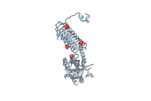
Organism: Homo sapiens
Method: X-RAY DIFFRACTION Resolution:1.80 Å Release Date: 2000-10-11 Classification: SIGNALING PROTEIN |
 |
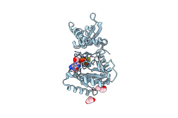
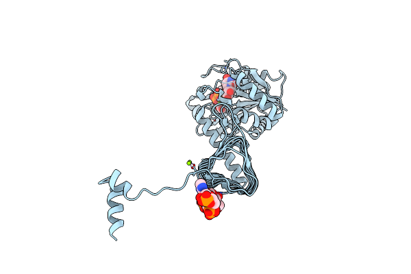
Human Guanylate Binding Protein-1 In Complex With The Gtp Analogue, Gmppnp.
Organism: Homo sapiens
Method: X-RAY DIFFRACTION Resolution:1.70 Å Release Date: 2000-09-27 Classification: SIGNALING PROTEIN Ligands: MG, GNP |