
Search Count: 12
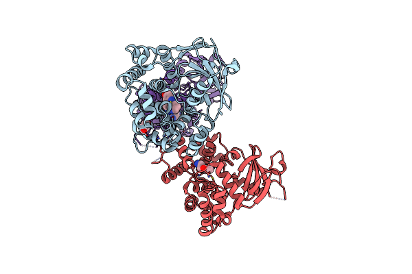 |
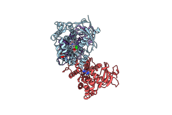
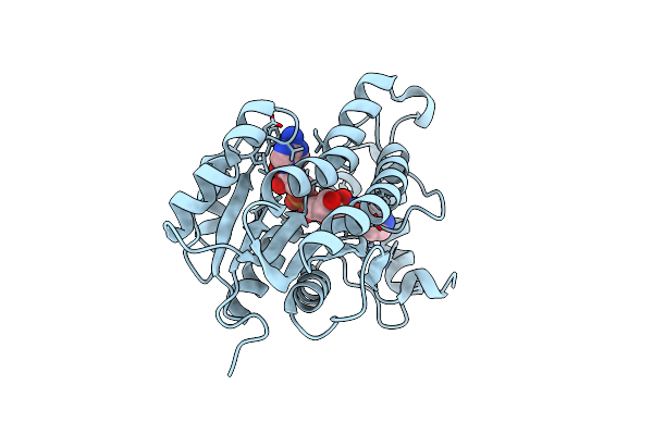
Structure Of The Mouse 8-Oxoguanine Dna Glycosylase Mogg1 In Complex With Ligand Th13579
Organism: Mus musculus
Method: X-RAY DIFFRACTION Release Date: 2025-06-25 Classification: DNA BINDING PROTEIN Ligands: A1ID3, GOL, NI |
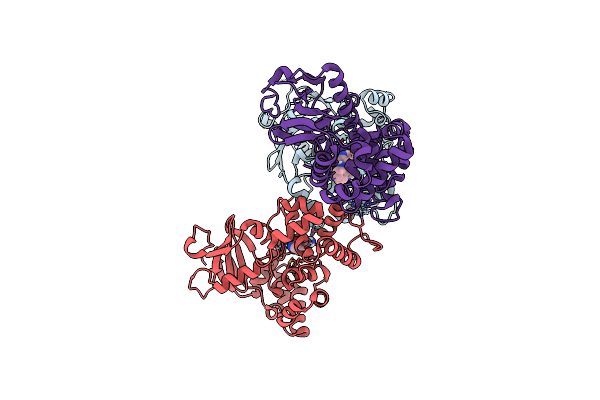 |
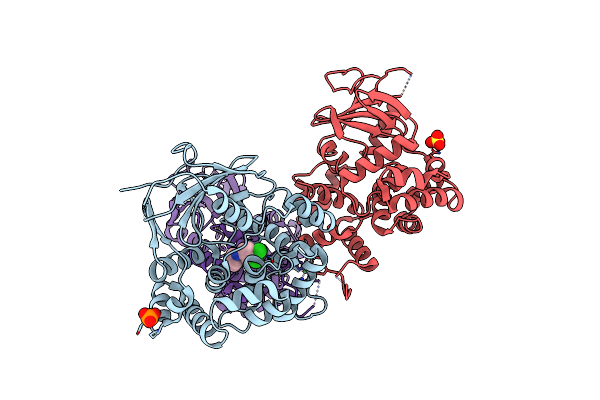
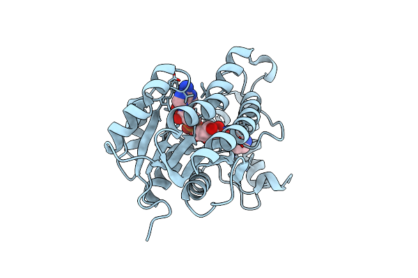
Structure Of The Mouse 8-Oxoguanine Dna Glycosylase Mogg1 In Complex With Ligand Th12163
Organism: Mus musculus
Method: X-RAY DIFFRACTION Release Date: 2025-06-25 Classification: DNA BINDING PROTEIN Ligands: A1IEJ, NI |
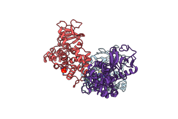 |
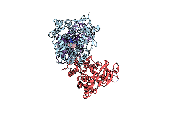
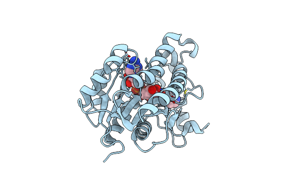
Structure Of The Mouse 8-Oxoguanine Dna Glycosylase Mogg1 In Complex With Ligand Th7399
Organism: Mus musculus
Method: X-RAY DIFFRACTION Release Date: 2025-05-21 Classification: DNA BINDING PROTEIN Ligands: A1IBK, NI, SO4 |
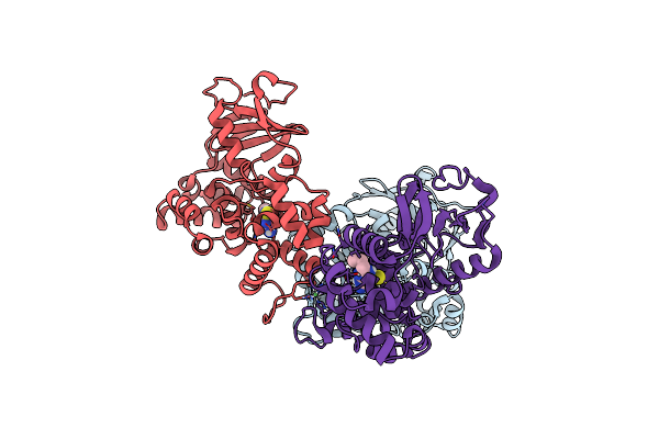 |
Structure Of The Mouse 8-Oxoguanine Dna Glycosylase Mogg1 In Complex With Ligand Th7420
Organism: Mus musculus
Method: X-RAY DIFFRACTION Release Date: 2025-05-21 Classification: DNA BINDING PROTEIN Ligands: A1IBL, NI |
 |
Structure Of The Mouse 8-Oxoguanine Dna Glycosylase Mogg1 In Complex With Ligand Th13677
Organism: Mus musculus
Method: X-RAY DIFFRACTION Release Date: 2025-05-21 Classification: DNA BINDING PROTEIN Ligands: A1IBJ, NI, SO4 |
 |
Structure Of The Mouse 8-Oxoguanine Dna Glycosylase Mogg1 In Complex With Ligand Th14445
Organism: Mus musculus
Method: X-RAY DIFFRACTION Release Date: 2025-05-21 Classification: DNA BINDING PROTEIN Ligands: A1IVU, PO4, NI, SO4 |
 |
Structure Of The Mouse 8-Oxoguanine Dna Glycosylase Mogg1 In Complex With Ligand Th2829
Organism: Mus musculus
Method: X-RAY DIFFRACTION Release Date: 2023-11-29 Classification: DNA BINDING PROTEIN Ligands: R4U, NI, GOL |
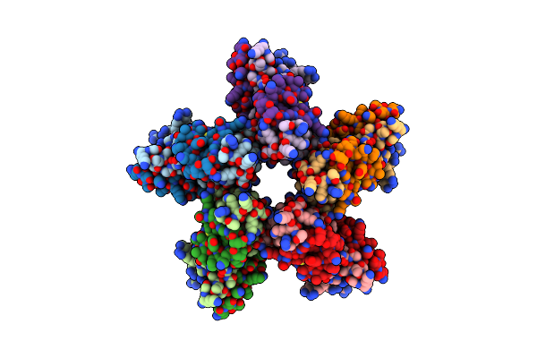 |
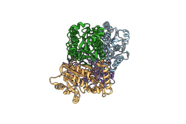
Fitting Of The H2A-H2B Histones In The Electron Microscopy Map Of The Complex Nucleoplasmin:H2A-H2B Histones (1:5).
Organism: Gallus gallus
Method: ELECTRON MICROSCOPY Resolution:19.50 Å Release Date: 2010-11-03 Classification: NUCLEAR PROTEIN |
 |
Crystal Structure Of Wild Type Enoyl-Acp(Coa) Reductase From Mycobacterium Tuberculosis In Complex With Nadh-Inh
Organism: Mycobacterium tuberculosis
Method: X-RAY DIFFRACTION Resolution:2.00 Å Release Date: 2007-07-24 Classification: OXIDOREDUCTASE Ligands: ZID |
 |
Crystal Structure Of Isoniazid-Resistant I21V Enoyl-Acp(Coa) Reductase Mutant Enzyme From Mycobacterium Tuberculosis In Complex With Nadh-Inh
Organism: Mycobacterium tuberculosis
Method: X-RAY DIFFRACTION Resolution:2.20 Å Release Date: 2007-07-24 Classification: OXIDOREDUCTASE Ligands: ZID |
 |
Crystal Structure Of Isoniazid-Resistant S94A Enoyl-Acp(Coa) Reductase Mutant Enzyme From Mycobacterium Tuberculosis In Complex With Nadh-Inh
Organism: Mycobacterium tuberculosis
Method: X-RAY DIFFRACTION Resolution:2.20 Å Release Date: 2007-07-24 Classification: OXIDOREDUCTASE Ligands: ZID |
 |
Crystal Structure Of Isoniazid-Resistant S94A Enoyl-Acp(Coa) Reductase Mutant Enzyme From Mycobacterium Tuberculosis Uncomplexed
Organism: Mycobacterium tuberculosis
Method: X-RAY DIFFRACTION Resolution:2.14 Å Release Date: 2007-07-24 Classification: OXIDOREDUCTASE |