
Search Count: 17
 |
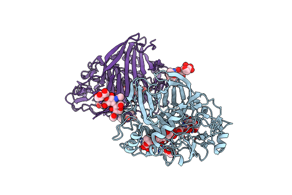
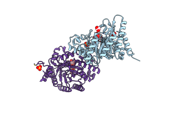
Organism: Myceliophthora thermophila
Method: X-RAY DIFFRACTION Resolution:2.60 Å Release Date: 2020-03-18 Classification: OXIDOREDUCTASE Ligands: FAD, NAG, SO4, CA, B3P, EDO, 144 |
 |
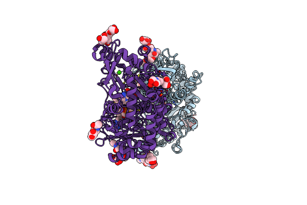
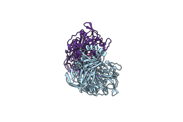
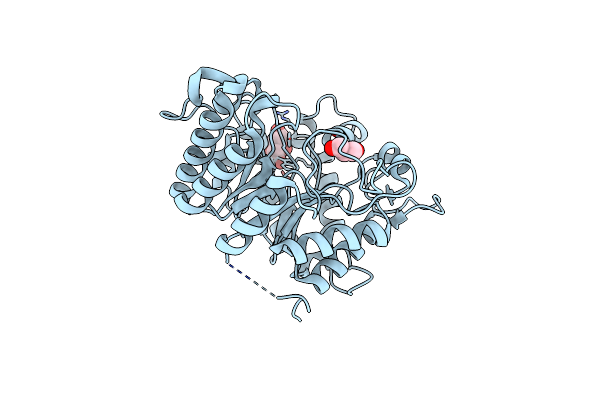
Biochemical And Structural Insights Into The Catalytic Mechanism Of Thermostable Cellobiohydrolase Cel7A From Industrially Relevant Fungus Myceliophthora Thermophila
Organism: Myceliophthora thermophila
Method: X-RAY DIFFRACTION Resolution:2.31 Å Release Date: 2017-12-13 Classification: HYDROLASE Ligands: MAN, 144 |
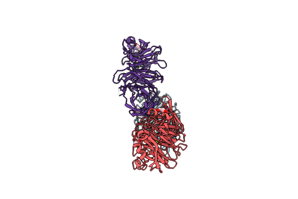 |
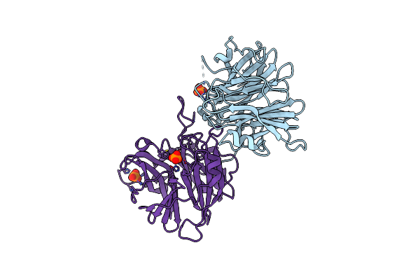
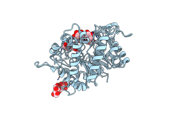
Crystal Structure Of Endo-1,5-Alpha-L-Arabinanase From Thermotoga Petrophila Rku-1
Organism: Thermotoga petrophila
Method: X-RAY DIFFRACTION Resolution:1.75 Å Release Date: 2014-02-05 Classification: HYDROLASE Ligands: PEG, CA |
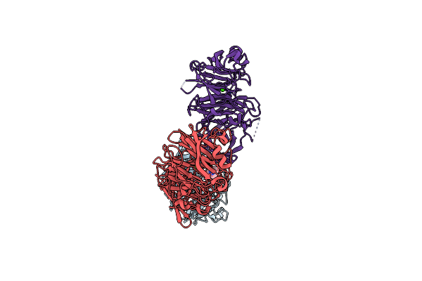 |
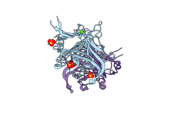
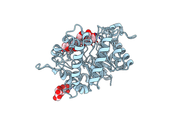
Crystal Structure Of Endo-1,5-Alpha-L-Arabinanase From Thermotoga Petrophila Rku-1 In Complex With Tris
Organism: Thermotoga petrophila
Method: X-RAY DIFFRACTION Resolution:1.76 Å Release Date: 2014-02-05 Classification: HYDROLASE Ligands: TRS, CA |
 |
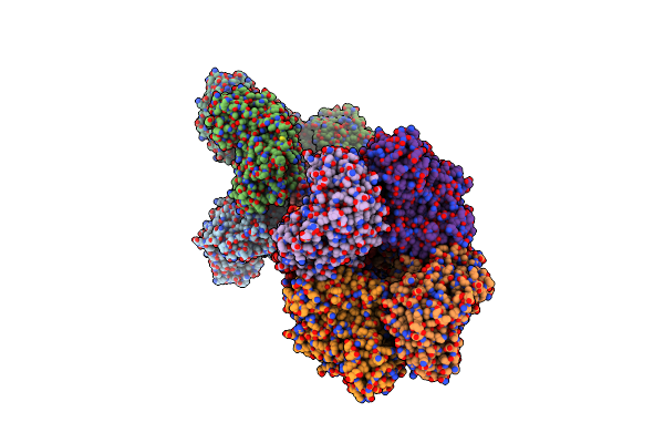
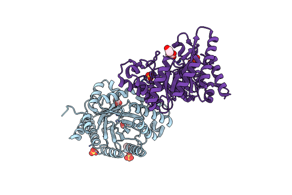
Crystal Structure Of Endo-1,5-Alpha-L-Arabinanase From A Bovine Ruminal Metagenomic Library
Organism: Bos taurus
Method: X-RAY DIFFRACTION Resolution:1.90 Å Release Date: 2014-02-05 Classification: HYDROLASE Ligands: IOD, GOL, NA |
 |
Crystal Structure Of Exo-1,5-Alpha-L-Arabinanase From Bovine Ruminal Metagenomic Library
Organism: Uncultured bacterium
Method: X-RAY DIFFRACTION Resolution:2.90 Å Release Date: 2014-02-05 Classification: HYDROLASE Ligands: PO4 |
 |
Structure Of The Catalytic Domain Of An Endo-1,3-Beta-Glucanase (Laminarinase) From Thermotoga Petrophila Rku-1
Organism: Thermotoga petrophila
Method: X-RAY DIFFRACTION Resolution:3.75 Å Release Date: 2012-03-14 Classification: HYDROLASE Ligands: CA, SO4 |
 |
Structure Of The Thermostable Gh51 Alpha-L-Arabinofuranosidase From Thermotoga Petrophila Rku-1
Organism: Thermotoga petrophila
Method: X-RAY DIFFRACTION Resolution:3.00 Å Release Date: 2012-02-01 Classification: HYDROLASE |
 |
Native Structure Of Endo-1,4-Beta-D-Mannanase From Thermotoga Petrophila Rku-1
Organism: Thermotoga petrophila
Method: X-RAY DIFFRACTION Resolution:1.42 Å Release Date: 2011-12-28 Classification: HYDROLASE |
 |
I222 Crystal Form Of The Hyperthermostable Endo-1,4-Beta-D-Mannanase From Thermotoga Petrophila Rku-1
Organism: Thermotoga petrophila
Method: X-RAY DIFFRACTION Resolution:1.40 Å Release Date: 2011-12-28 Classification: HYDROLASE Ligands: TRS, GOL, PO4 |
 |
Structure Of The Hyperthermostable Endo-1,4-Beta-D-Mannanase From Thermotoga Petrophila Rku-1 In Complex With Beta-D-Glucose
Organism: Thermotoga petrophila
Method: X-RAY DIFFRACTION Resolution:1.55 Å Release Date: 2011-12-28 Classification: HYDROLASE Ligands: BGC |
 |
Structure Of The Hyperthermostable Endo-1,4-Beta-D-Mannanase From Thermotoga Petrophila Rku-1 With Three Glycerol Molecules
Organism: Thermotoga petrophila rku-1
Method: X-RAY DIFFRACTION Resolution:1.50 Å Release Date: 2011-12-28 Classification: HYDROLASE Ligands: GOL, TRS |
 |
Structure Of The Hyperthermostable Endo-1,4-Beta-D-Mannanase From Thermotoga Petrophila Rku-1 With Citrate And Glycerol
Organism: Thermotoga petrophila rku-1
Method: X-RAY DIFFRACTION Resolution:1.50 Å Release Date: 2011-12-28 Classification: HYDROLASE Ligands: CIT, GOL |
 |
Structure Of The Hyperthermostable Endo-1,4-Beta-D-Mannanase From Thermotoga Petrophila Rku-1 In Complex With Three Maltose Molecules
Organism: Thermotoga petrophila rku-1
Method: X-RAY DIFFRACTION Resolution:1.55 Å Release Date: 2011-12-28 Classification: HYDROLASE Ligands: GOL |
 |
Structure Of The Hyperthermostable Endo-1,4-Beta-D-Mannanase From Thermotoga Petrophila Rku-1 With Maltose And Glycerol
Organism: Thermotoga petrophila rku-1
Method: X-RAY DIFFRACTION Resolution:1.92 Å Release Date: 2011-12-28 Classification: HYDROLASE |
 |
Organism: Thermotoga petrophila rku-1
Method: X-RAY DIFFRACTION Resolution:1.58 Å Release Date: 2011-05-04 Classification: HYDROLASE Ligands: SO4, ACT |
 |
Crystal Structure Of Xylanase 10B From Thermotoga Petrophila Rku-1 In Complex With Xylobiose
Organism: Thermotoga petrophila rku-1
Method: X-RAY DIFFRACTION Resolution:1.88 Å Release Date: 2011-05-04 Classification: HYDROLASE Ligands: ACT, SO4 |