
Search Count: 18
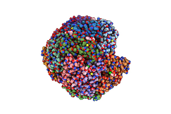 |
Crystal Structure Of Structure Of Wt Bfrb From Pseudomonas Aeruginosa In Complex With A Protein-Protein Interaction Inhibitor Km-5-25
Organism: Pseudomonas aeruginosa pao1
Method: X-RAY DIFFRACTION Release Date: 2025-06-18 Classification: OXIDOREDUCTASE Ligands: K, HEM, PG4, A1BYB |
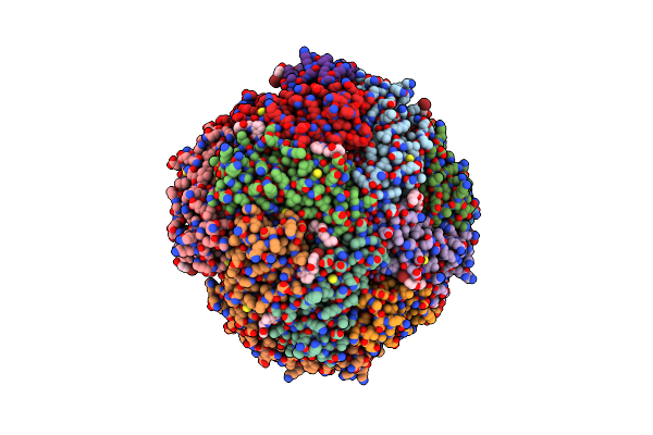 |
Crystal Structure Of Structure Of Wt Bfrb From Pseudomonas Aeruginosa In Complex With A Protein-Protein Interaction Inhibitor Km-5-35
Organism: Pseudomonas aeruginosa pao1
Method: X-RAY DIFFRACTION Release Date: 2025-06-18 Classification: OXIDOREDUCTASE Ligands: HEM, A1BYC, K, NA, PG4 |
 |
Organism: Enterobacteria phage t4, Homo sapiens, Mus musculus
Method: X-RAY DIFFRACTION Resolution:7.70 Å Release Date: 2016-03-23 Classification: SIGNALING PROTEIN |
 |
Crystal Structure Of Rhodopsin Bound To Arrestin By Femtosecond X-Ray Laser
Organism: Enterobacteria phage t4, Homo sapiens, Mus musculus
Method: X-RAY DIFFRACTION Resolution:3.30 Å Release Date: 2015-07-29 Classification: SIGNALING PROTEIN |
 |
Organism: Escherichia coli o104:h4, Mus musculus
Method: X-RAY DIFFRACTION Resolution:2.80 Å Release Date: 2014-01-29 Classification: TRANSCRIPTION |
 |
Organism: Arabidopsis thaliana
Method: X-RAY DIFFRACTION Resolution:2.60 Å Release Date: 2012-02-15 Classification: SIGNALING PROTEIN Ligands: SO4, MG |
 |
Organism: Arabidopsis thaliana
Method: X-RAY DIFFRACTION Resolution:1.90 Å Release Date: 2012-02-15 Classification: HYDROLASE Ligands: MG |
 |
Crystal Structure Of Abscisic Acid Bound Pyl2 In Complex With Type 2C Protein Phosphatase Abi2
Organism: Arabidopsis thaliana
Method: X-RAY DIFFRACTION Resolution:2.50 Å Release Date: 2012-02-15 Classification: SIGNALING PROTEIN Ligands: A8S, MG |
 |
Organism: Arabidopsis thaliana
Method: X-RAY DIFFRACTION Resolution:1.90 Å Release Date: 2011-12-14 Classification: TRANSFERASE Ligands: CO |
 |
Organism: Arabidopsis thaliana
Method: X-RAY DIFFRACTION Resolution:2.30 Å Release Date: 2011-12-14 Classification: TRANSFERASE |
 |
Crystal Structure Of The Ligand Binding Domain Of Human Testicular Receptor 4
Organism: Homo sapiens
Method: X-RAY DIFFRACTION Resolution:3.00 Å Release Date: 2010-11-10 Classification: SIGNALING PROTEIN |
 |
Organism: Arabidopsis thaliana
Method: X-RAY DIFFRACTION Resolution:1.85 Å Release Date: 2010-08-25 Classification: hormone binding protein Ligands: PYV, EPE |
 |
Crystal Structure Of Pyrabactin-Bound Abscisic Acid Receptor Pyl1 In Complex With Type 2C Protein Phosphatase Abi1
Organism: Arabidopsis thaliana
Method: X-RAY DIFFRACTION Resolution:2.15 Å Release Date: 2010-08-25 Classification: PROTEIN BINDING Ligands: PYV, MG |
 |
Crystal Structure Of The Abscisic Receptor Pyl2 Mutant A93F In Complex With Pyrabactin
Organism: Arabidopsis thaliana
Method: X-RAY DIFFRACTION Resolution:2.10 Å Release Date: 2010-08-25 Classification: hormone binding protein Ligands: PYV |
 |
Crystal Structure Of Pyrabactin Bound Abscisic Acid Receptor Pyl2 Mutant A93F In Complex With Type 2C Protein Phosphatase Hab1
Organism: Arabidopsis thaliana
Method: X-RAY DIFFRACTION Resolution:2.56 Å Release Date: 2010-08-25 Classification: PROTEIN BINDING Ligands: PYV, MG, SO4 |
 |
Crystal Structure Of Pyrabactin-Bound Abscisic Acid Receptor Pyl2 Mutant A93F In Complex With Type 2C Protein Phosphatase Abi2
Organism: Arabidopsis thaliana
Method: X-RAY DIFFRACTION Resolution:2.10 Å Release Date: 2010-08-25 Classification: PROTEIN BINDING Ligands: PYV, MG |
 |
Crystal Structure Of Dht-Bound Androgen Receptor In Complex With The First Motif Of Steroid Receptor Coactivator 3
Organism: Homo sapiens
Method: X-RAY DIFFRACTION Resolution:1.55 Å Release Date: 2010-01-12 Classification: PROTEIN BINDING Ligands: DHT |
 |
Crystal Structure Of Dht-Bound Androgen Receptor In Complex With The Third Motif Of Steroid Receptor Coactivator 3
Organism: Homo sapiens
Method: X-RAY DIFFRACTION Resolution:2.00 Å Release Date: 2010-01-12 Classification: PROTEIN BINDING Ligands: DHT |