
Search Count: 42
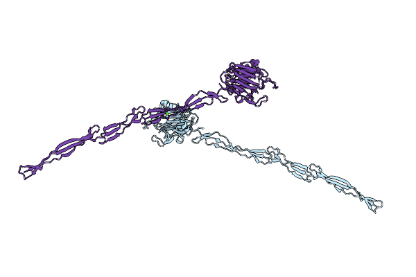 |
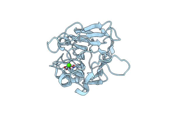
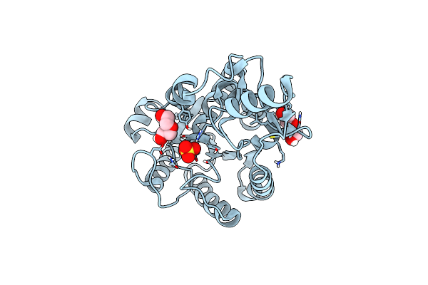
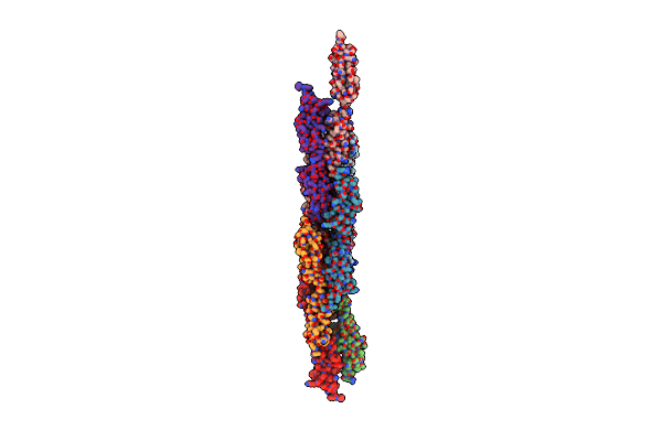
Structure Of Aap A Domain And B-Repeats (Residues 351-813) From Staphylococcus Epidermidis
Organism: Staphylococcus epidermidis rp62a
Method: X-RAY DIFFRACTION Resolution:2.30 Å Release Date: 2023-05-03 Classification: CELL ADHESION Ligands: CA, CL |
 |
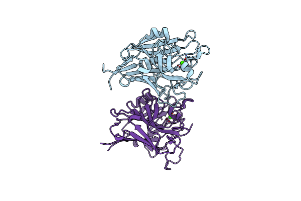
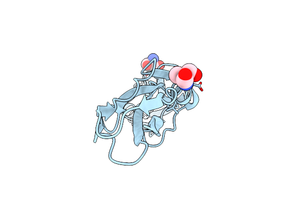
Structure Of Pls A-Domain (Residues 391-656; 513-518 Deletion Mutant) From Staphylococcus Aureus
Organism: Staphylococcus aureus
Method: X-RAY DIFFRACTION Resolution:2.75 Å Release Date: 2022-11-09 Classification: CELL ADHESION Ligands: CA |
 |
Organism: Staphylococcus aureus (strain nctc 8325 / ps 47)
Method: X-RAY DIFFRACTION Resolution:1.65 Å Release Date: 2022-11-02 Classification: CELL ADHESION Ligands: CA, EDO |
 |
Organism: Staphylococcus aureus subsp. aureus nctc 8325
Method: X-RAY DIFFRACTION Resolution:1.21 Å Release Date: 2022-10-26 Classification: CELL ADHESION Ligands: CA |
 |
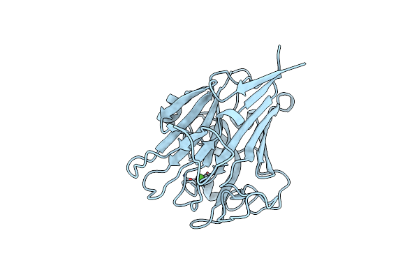
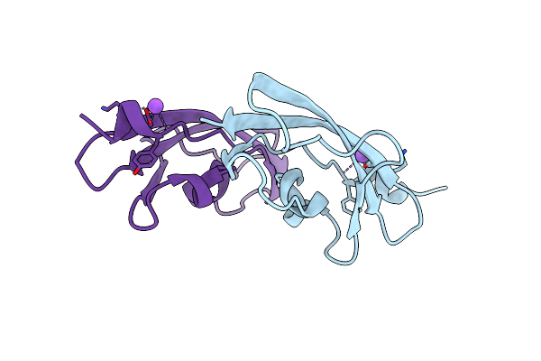
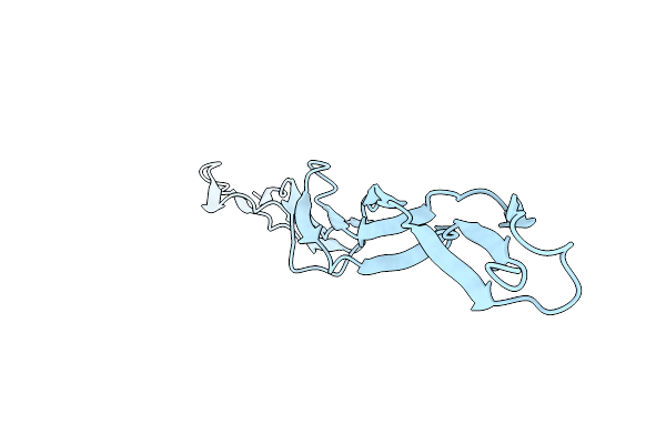
Structure Of Aap A-Domain (Residues 351-605) From Staphylococcus Epidermidis
Organism: Staphylococcus epidermidis (strain atcc 35984 / rp62a)
Method: X-RAY DIFFRACTION Resolution:1.30 Å Release Date: 2022-10-19 Classification: CELL ADHESION Ligands: CA, CL |
 |
Streptococcal High Identity Repeats In Tandem (Shirt) Domains 3-4 From Cell Surface Protein Sgo_0707
Organism: Streptococcus gordonii (strain challis / atcc 35105 / bcrc 15272 / ch1 / dl1 / v288)
Method: X-RAY DIFFRACTION Resolution:1.35 Å Release Date: 2021-06-09 Classification: CELL ADHESION Ligands: CL, TRS |
 |
Streptococcal High Identity Repeats In Tandem (Shirt) Domain 2 From Cell Surface Protein Sgo_0707
Organism: Streptococcus gordonii (strain challis / atcc 35105 / bcrc 15272 / ch1 / dl1 / v288)
Method: X-RAY DIFFRACTION Resolution:0.95 Å Release Date: 2021-06-09 Classification: CELL ADHESION |
 |
Streptococcal High Identity Repeats In Tandem (Shirt) Domain 10 From Cell Surface Protein Sgo_0707
Organism: Streptococcus gordonii (strain challis / atcc 35105 / bcrc 15272 / ch1 / dl1 / v288)
Method: X-RAY DIFFRACTION Resolution:0.82 Å Release Date: 2021-06-09 Classification: CELL ADHESION Ligands: IS8 |
 |
Structure Of Salmonella Ser. Paratyphi A Lipopolysaccharide Acetyltransferase Periplasmic Domain
Organism: Salmonella paratyphi a (strain atcc 9150 / sarb42)
Method: X-RAY DIFFRACTION Resolution:1.08 Å Release Date: 2020-08-26 Classification: TRANSFERASE Ligands: SO4, PEG |
 |
Organism: Lactobacillus acidophilus (strain atcc 700396 / nck56 / n2 / ncfm)
Method: X-RAY DIFFRACTION Resolution:1.25 Å Release Date: 2019-12-18 Classification: STRUCTURAL PROTEIN |
 |
Organism: Lactobacillus acidophilus
Method: X-RAY DIFFRACTION Resolution:1.07 Å Release Date: 2019-12-11 Classification: STRUCTURAL PROTEIN Ligands: SO4, NA |
 |
Structure Of Ribr, The Most N-Terminal Rib Domain From Group B Streptococcus Species Streptococcus Agalactiae
Organism: Streptococcus agalactiae
Method: X-RAY DIFFRACTION Resolution:1.70 Å Release Date: 2019-12-11 Classification: STRUCTURAL PROTEIN |
 |
Structure Of Tandemly Arrayed Consecutive Rib Domains (Rib2R) From Group B Streptococcal Species Streptococcus Agalactiae
Organism: Streptococcus agalactiae
Method: X-RAY DIFFRACTION Resolution:2.30 Å Release Date: 2019-12-11 Classification: STRUCTURAL PROTEIN |
 |
Organism: Streptococcus pyogenes
Method: X-RAY DIFFRACTION Resolution:1.80 Å Release Date: 2019-12-11 Classification: STRUCTURAL PROTEIN |
 |
Organism: Staphylococcus aureus
Method: X-RAY DIFFRACTION Resolution:1.60 Å Release Date: 2016-09-28 Classification: STRUCTURAL PROTEIN |
 |
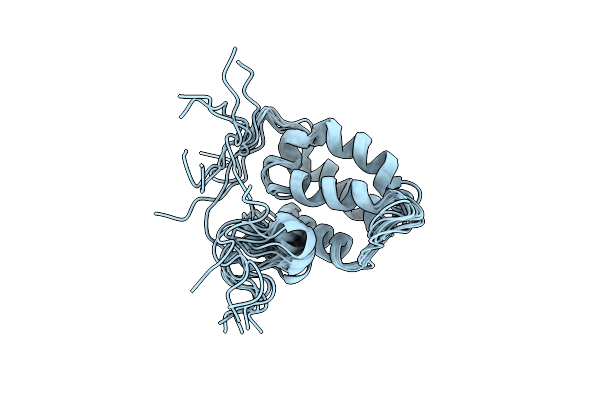
Organism: Streptomyces coelicolor
Method: SOLUTION NMR Release Date: 2016-08-03 Classification: TRANSCRIPTION Ligands: ZN |
 |
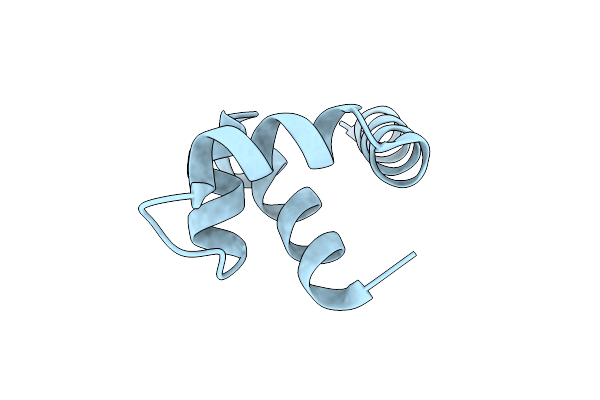
Organism: Streptomyces coelicolor
Method: SOLUTION NMR Release Date: 2016-08-03 Classification: TRANSCRIPTION |
 |
Organism: Bacillus phage sf6
Method: X-RAY DIFFRACTION Resolution:1.40 Å Release Date: 2015-12-30 Classification: VIRAL PROTEIN |
 |
Organism: Staphylococcus aureus
Method: X-RAY DIFFRACTION Resolution:1.60 Å Release Date: 2015-06-03 Classification: STRUCTURAL PROTEIN Ligands: CL |
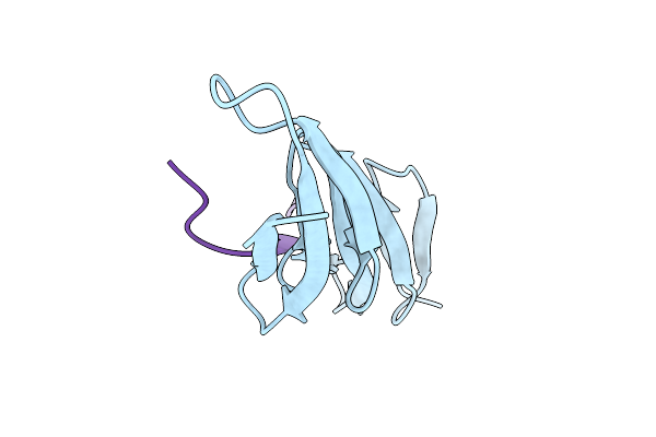 |
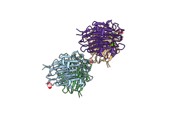
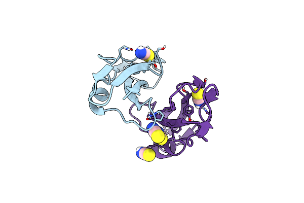
Crystal Structure Of The Second And Third Fibronectin F1 Modules In Complex With A Fragment Of Bbk32 From Borrelia Burgdorferi
Organism: Homo sapiens, Borrelia burgdorferi
Method: X-RAY DIFFRACTION Resolution:1.96 Å Release Date: 2014-07-02 Classification: CELL ADHESION |