
Search Count: 21
 |
Organism: Homo sapiens
Method: SOLUTION NMR Release Date: 2019-01-02 Classification: LIPID BINDING PROTEIN |
 |
Organism: Homo sapiens
Method: SOLUTION NMR Release Date: 2018-12-26 Classification: LIPID BINDING PROTEIN |
 |
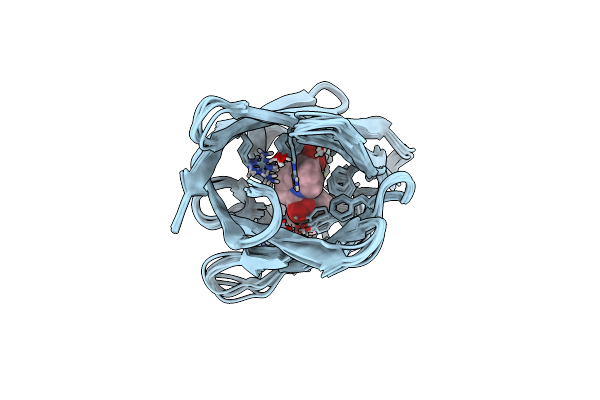
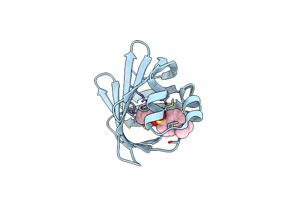
Organism: Homo sapiens
Method: SOLUTION NMR Release Date: 2018-12-26 Classification: LIPID BINDING PROTEIN Ligands: 2VN |
 |
Organism: Homo sapiens
Method: SOLUTION NMR Release Date: 2014-10-29 Classification: LIPID BINDING PROTEIN, TRANSPORT PROTEIN Ligands: KTR |
 |
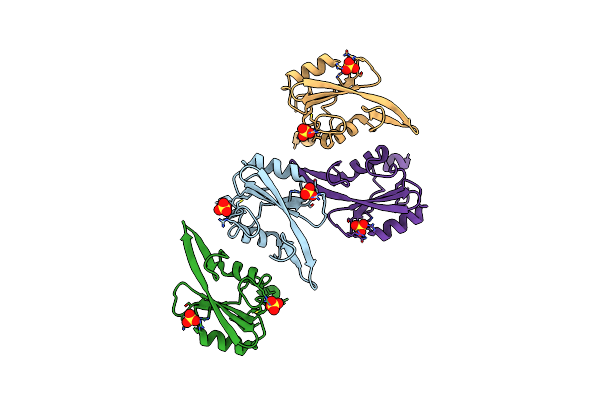
Organism: Clostridium perfringens
Method: X-RAY DIFFRACTION Resolution:1.80 Å Release Date: 2013-03-20 Classification: TOXIN Ligands: MG, EDO |
 |
Organism: Clostridium perfringens
Method: X-RAY DIFFRACTION Resolution:1.80 Å Release Date: 2012-02-08 Classification: TRANSPORT PROTEIN Ligands: PEG |
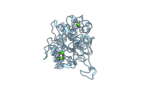 |
Crystal Structure Of The Basic Protease Bprv From The Ovine Footrot Pathogen, Dichelobacter Nodosus
Organism: Dichelobacter nodosus
Method: X-RAY DIFFRACTION Resolution:2.00 Å Release Date: 2011-10-19 Classification: HYDROLASE Ligands: CA |
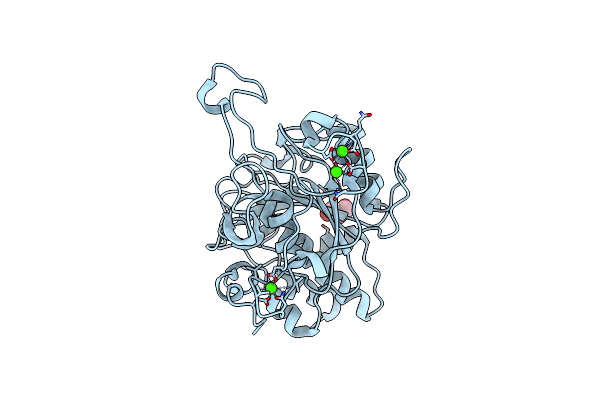 |
Crystal Structure Of The Basic Protease Bprb From The Ovine Footrot Pathogen, Dichelobacter Nodosus
Organism: Dichelobacter nodosus
Method: X-RAY DIFFRACTION Resolution:1.80 Å Release Date: 2011-10-19 Classification: HYDROLASE Ligands: CA, GOL, CL |
 |
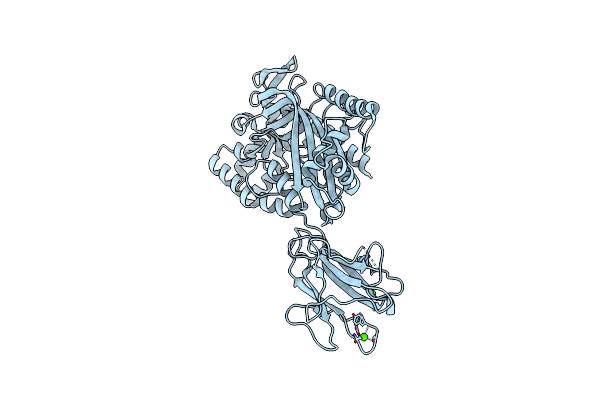
X-Ray Structure Of Ifabp From Human And Rat With Bound Fluorescent Fatty Acid Analogue
Organism: Homo sapiens
Method: X-RAY DIFFRACTION Resolution:1.90 Å Release Date: 2011-07-20 Classification: TRANSPORT PROTEIN Ligands: 11D, MG |
 |
X-Ray Structure Of Ifabp From Human And Rat With Bound Fluorescent Fatty Acid Analogue
Organism: Rattus norvegicus
Method: X-RAY DIFFRACTION Resolution:1.60 Å Release Date: 2011-07-20 Classification: TRANSPORT PROTEIN Ligands: 11D |
 |
Organism: Dichelobacter nodosus
Method: X-RAY DIFFRACTION Resolution:2.00 Å Release Date: 2010-12-08 Classification: HYDROLASE Ligands: CA |
 |
Organism: Dichelobacter nodosus
Method: X-RAY DIFFRACTION Resolution:1.70 Å Release Date: 2010-12-08 Classification: HYDROLASE Ligands: CA, ACT, PEG |
 |
Organism: Dichelobacter nodosus
Method: X-RAY DIFFRACTION Resolution:2.10 Å Release Date: 2010-12-08 Classification: HYDROLASE Ligands: CA |
 |
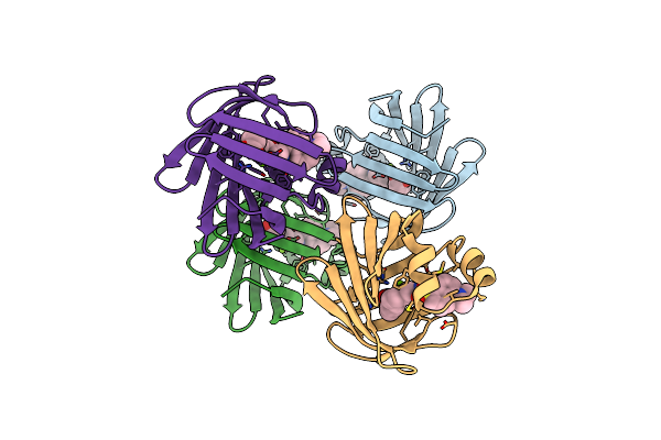
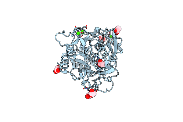
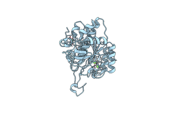
Organism: Plasmodium falciparum
Method: X-RAY DIFFRACTION Resolution:2.10 Å Release Date: 2009-01-27 Classification: HYDROLASE Ligands: ZN, MG, GOL |
 |
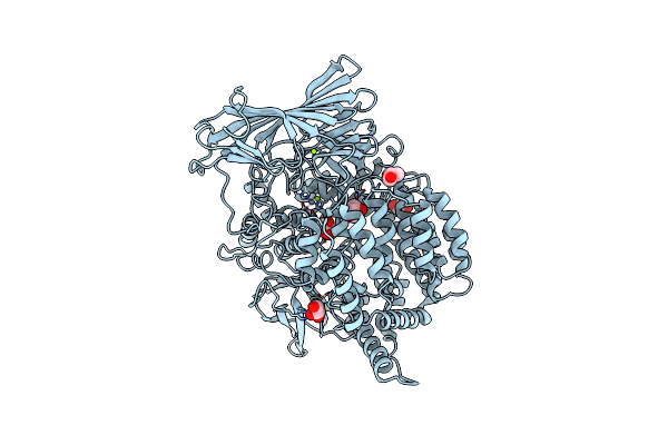
Structure Of The M1 Alanylaminopeptidase From Malaria Complexed With Bestatin
Organism: Plasmodium falciparum
Method: X-RAY DIFFRACTION Resolution:1.65 Å Release Date: 2009-01-27 Classification: HYDROLASE INHIBITOR Ligands: ZN, BES, GOL, MG |
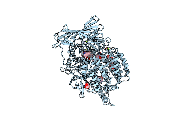 |
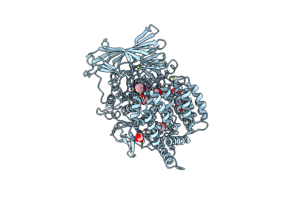
Structure Of The M1 Alanylaminopeptidase From Malaria Complexed With The Phosphinate Dipeptide Analog
Organism: Plasmodium falciparum
Method: X-RAY DIFFRACTION Resolution:2.00 Å Release Date: 2009-01-27 Classification: HYDROLASE INHIBITOR Ligands: ZN, BEY, GOL, MG |
 |
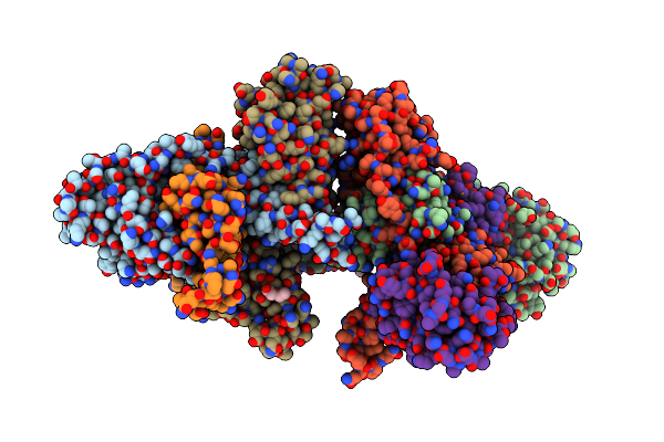
Organism: Homo sapiens
Method: X-RAY DIFFRACTION Resolution:2.10 Å Release Date: 2008-07-22 Classification: SIGNALING PROTEIN Ligands: SO4 |
 |
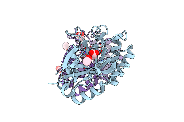
Organism: Bacillus subtilis
Method: X-RAY DIFFRACTION Resolution:2.50 Å Release Date: 2008-02-26 Classification: REPLICATION/DNA |
 |
Organism: Photorhabdus luminescens subsp. laumondii
Method: X-RAY DIFFRACTION Resolution:2.00 Å Release Date: 2007-09-04 Classification: UNKNOWN FUNCTION Ligands: CA |
 |
Organism: Unidentified phage
Method: X-RAY DIFFRACTION Resolution:1.60 Å Release Date: 2006-12-05 Classification: HYDROLASE Ligands: ACT |