
Search Count: 34
 |
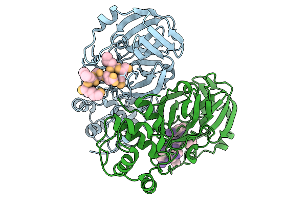
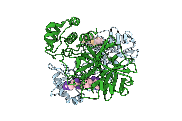
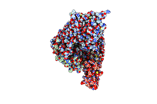
Organism: Porcine epidemic diarrhea virus
Method: X-RAY DIFFRACTION Resolution:2.23 Å Release Date: 2026-01-14 Classification: VIRAL PROTEIN |
 |
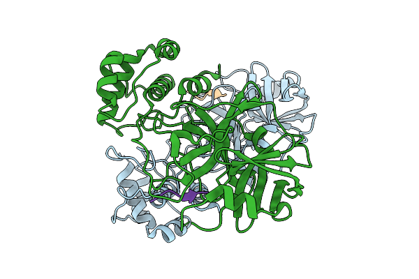
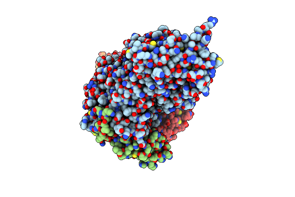
Organism: Porcine epidemic diarrhea virus
Method: X-RAY DIFFRACTION Resolution:2.30 Å Release Date: 2025-12-31 Classification: VIRAL PROTEIN |
 |
Organism: Porcine epidemic diarrhea virus
Method: X-RAY DIFFRACTION Resolution:2.31 Å Release Date: 2025-12-31 Classification: VIRAL PROTEIN |
 |
Organism: Porcine epidemic diarrhea virus
Method: X-RAY DIFFRACTION Resolution:1.96 Å Release Date: 2025-12-24 Classification: VIRAL PROTEIN |
 |
Organism: Porcine epidemic diarrhea virus
Method: X-RAY DIFFRACTION Resolution:1.93 Å Release Date: 2025-12-17 Classification: VIRAL PROTEIN |
 |
Organism: Porcine epidemic diarrhea virus
Method: X-RAY DIFFRACTION Resolution:1.78 Å Release Date: 2025-12-17 Classification: VIRAL PROTEIN |
 |
Organism: Porcine epidemic diarrhea virus
Method: X-RAY DIFFRACTION Resolution:1.88 Å Release Date: 2025-12-17 Classification: VIRAL PROTEIN |
 |
Organism: Porcine epidemic diarrhea virus
Method: X-RAY DIFFRACTION Resolution:1.95 Å Release Date: 2025-10-22 Classification: VIRAL PROTEIN |
 |
Organism: Porcine epidemic diarrhea virus
Method: X-RAY DIFFRACTION Resolution:1.84 Å Release Date: 2025-01-15 Classification: VIRAL PROTEIN |
 |
Organism: Porcine epidemic diarrhea virus
Method: X-RAY DIFFRACTION Resolution:2.88 Å Release Date: 2024-10-16 Classification: VIRAL PROTEIN |
 |
Organism: Porcine epidemic diarrhea virus, Synthetic construct
Method: ELECTRON MICROSCOPY Release Date: 2023-11-15 Classification: REPLICATION, TRANSFERASE/RNA Ligands: ZN |
 |
Organism: Porcine epidemic diarrhea virus, Synthetic construct
Method: ELECTRON MICROSCOPY Release Date: 2023-03-29 Classification: REPLICATION Ligands: ZN |
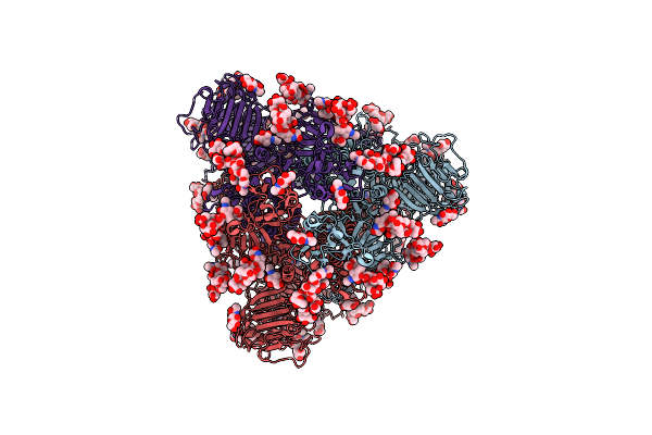 |
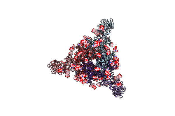
Cryo-Em Map Of Pedv (Pintung 52) S Protein With All Three Protomers In The D0-Down Conformation Determined In Situ On Intact Viral Particles.
Organism: Porcine epidemic diarrhea virus
Method: ELECTRON MICROSCOPY Release Date: 2022-08-03 Classification: VIRAL PROTEIN Ligands: NAG |
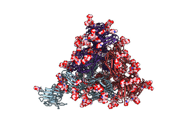 |
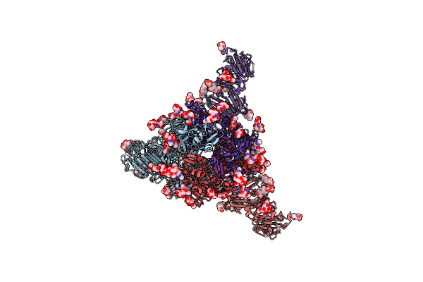
Cryo-Em Map Of Pedv S Protein With One Protomer In The D0-Up Conformation While The Other Two In The D0-Down Conformation
Organism: Porcine epidemic diarrhea virus
Method: ELECTRON MICROSCOPY Release Date: 2022-08-03 Classification: VIRAL PROTEIN Ligands: NAG |
 |
Organism: Porcine epidemic diarrhea virus
Method: ELECTRON MICROSCOPY Release Date: 2022-08-03 Classification: VIRAL PROTEIN Ligands: NAG |
 |
Cryo-Em Map Of Ipec-J2 Cell-Derived Pedv Pt52 S Protein One D0-Down And Two D0-Up
Organism: Porcine epidemic diarrhea virus
Method: ELECTRON MICROSCOPY Release Date: 2022-08-03 Classification: VIRAL PROTEIN Ligands: NAG |
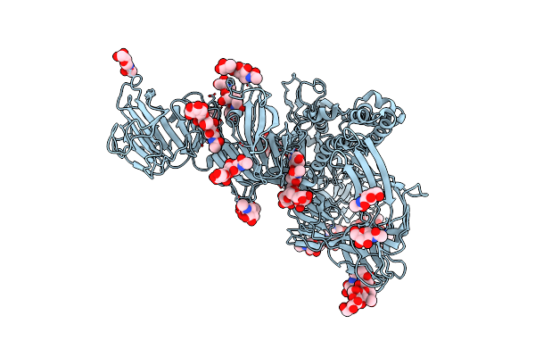 |
Symmetry-Expanded And Locally Refined Protomer Structure Of Ipec-J2 Cell-Derived Pedv Pt52 S With A Ctd-Close Conformation
Organism: Porcine epidemic diarrhea virus
Method: ELECTRON MICROSCOPY Release Date: 2022-08-03 Classification: VIRAL PROTEIN Ligands: NAG |
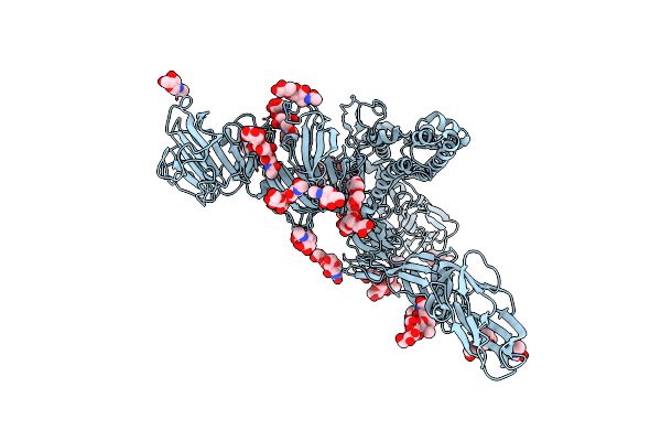 |
Symmetry-Expanded And Locally Refined Protomer Structure Of Ipec-J2 Cell-Derived Pedv Pt52 S With A Ctd-Open Conformation
Organism: Porcine epidemic diarrhea virus
Method: ELECTRON MICROSCOPY Resolution:3.30 Å Release Date: 2022-08-03 Classification: VIRAL PROTEIN Ligands: NAG |
 |
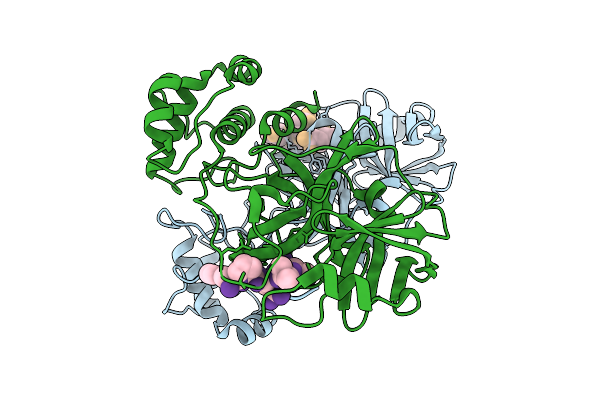
Porcine Epidemic Diarrhea Virus Papain-Like Protease 2 C44S Mutant In Complex With Mono Ubiquitin
Organism: Porcine epidemic diarrhea virus, Bos taurus
Method: X-RAY DIFFRACTION Resolution:2.20 Å Release Date: 2021-10-20 Classification: VIRAL PROTEIN/PROTEIN BINDING Ligands: ZN |
 |
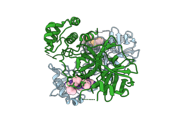
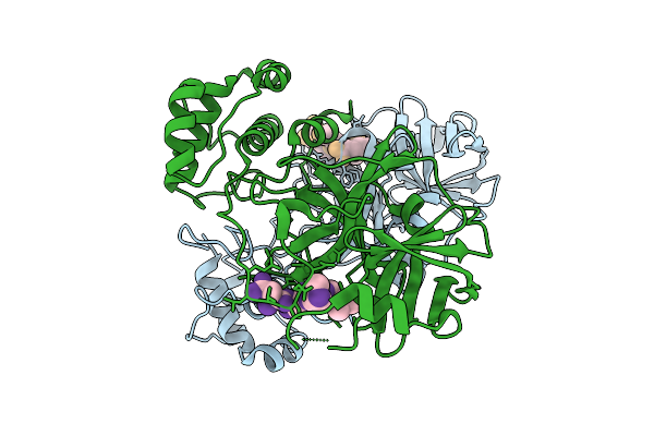
Organism: Porcine epidemic diarrhea virus, Homo sapiens
Method: X-RAY DIFFRACTION Resolution:3.10 Å Release Date: 2021-07-07 Classification: HYDROLASE Ligands: ZN, AYE |