
Search Count: 7
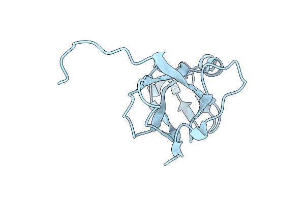 |
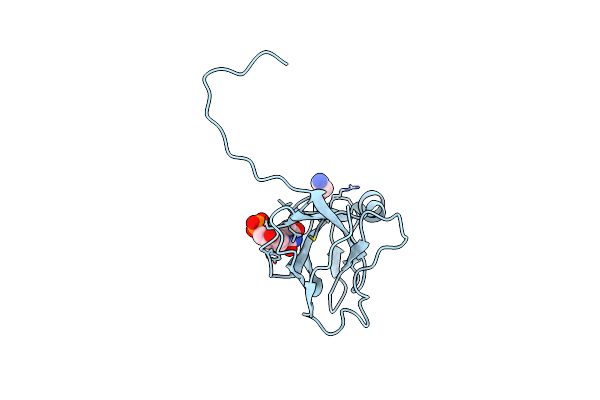
Organism: Mason-pfizer monkey virus
Method: X-RAY DIFFRACTION Resolution:1.85 Å Release Date: 2006-11-21 Classification: HYDROLASE |
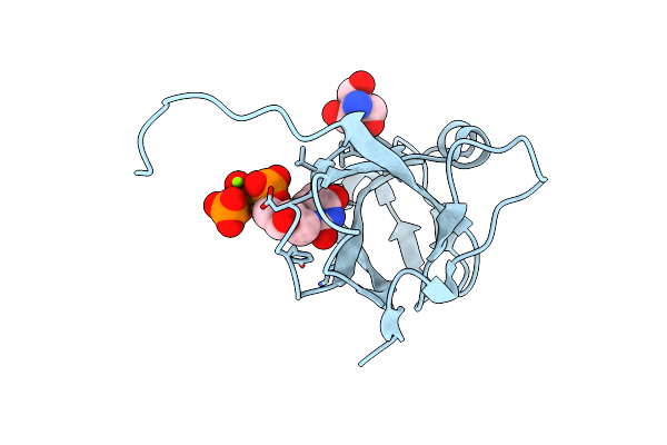 |
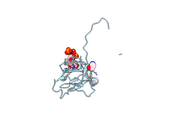
Crystal Structure Of M-Pmv Dutpase Complexed With Dupnpp, Substrate Analogue
Organism: Mason-pfizer monkey virus
Method: X-RAY DIFFRACTION Resolution:1.53 Å Release Date: 2006-11-21 Classification: HYDROLASE Ligands: MG, DUP, TRS |
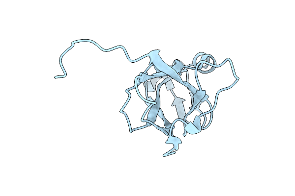 |
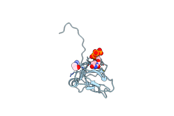
Organism: Mason-pfizer monkey virus
Method: X-RAY DIFFRACTION Resolution:1.70 Å Release Date: 2006-11-07 Classification: HYDROLASE |
 |
Organism: Escherichia coli
Method: X-RAY DIFFRACTION Resolution:1.93 Å Release Date: 2004-09-07 Classification: HYDROLASE Ligands: MG, ACT, DUP, TRS |
 |
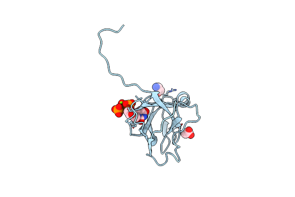
Crystal Structure Of Inactive Mutant Dutpase Complexed With Substrate Analogue Imido-Dutp
Organism: Escherichia coli
Method: X-RAY DIFFRACTION Resolution:1.70 Å Release Date: 2004-09-07 Classification: HYDROLASE Ligands: MG, DUP, TRS |
 |
Organism: Escherichia coli
Method: X-RAY DIFFRACTION Resolution:1.47 Å Release Date: 2004-09-07 Classification: HYDROLASE Ligands: UMP, TRS |
 |
Organism: Escherichia coli
Method: X-RAY DIFFRACTION Resolution:1.95 Å Release Date: 2004-09-07 Classification: HYDROLASE Ligands: MG, DUT, TRS |