
Search Count: 14
 |
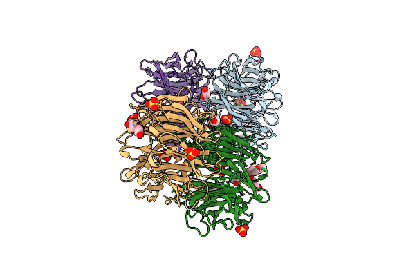
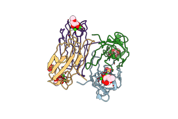
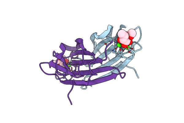
Crystal Structure Of Bp39L Lectin From Burkholderia Pseudomallei At 1.7 A Resolution
Organism: Burkholderia pseudomallei
Method: X-RAY DIFFRACTION Resolution:1.70 Å Release Date: 2019-12-04 Classification: SUGAR BINDING PROTEIN Ligands: SO4 |
 |
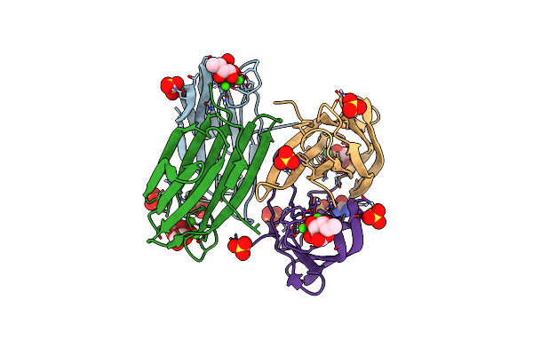
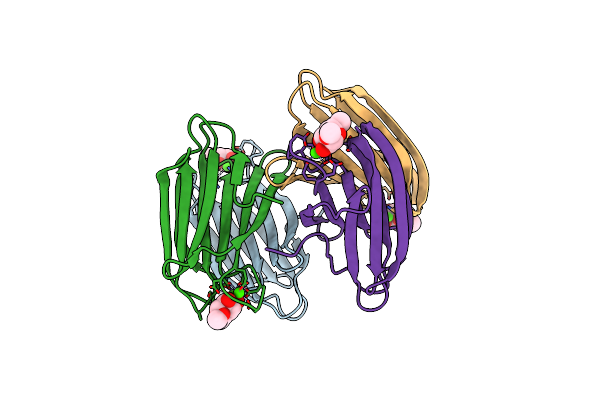
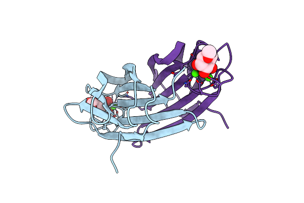
Crystal Structure Of Cv39L Lectin From Chromobacterium Violaceum At 1.8 A Resolution
Organism: Chromobacterium violaceum (strain atcc 12472 / dsm 30191 / jcm 1249 / nbrc 12614 / ncimb 9131 / nctc 9757)
Method: X-RAY DIFFRACTION Resolution:1.80 Å Release Date: 2019-12-04 Classification: SUGAR BINDING PROTEIN Ligands: SO4, EDO, PEG, 1PE |
 |
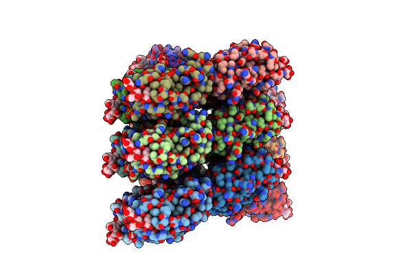
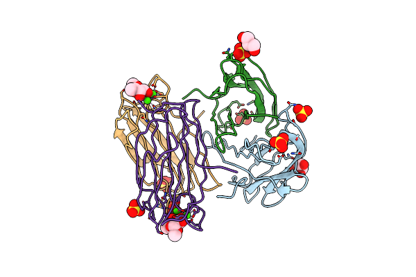
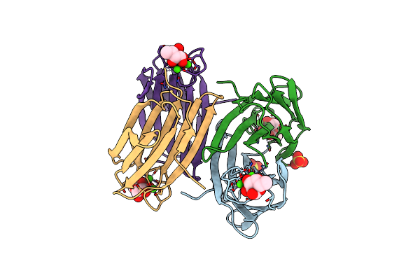
Perdeuterated Pseudomonas Aeruginosa Lectin Ii Complex With Hydrogenated L-Fucose And Calcium
Organism: Pseudomonas aeruginosa
Method: X-RAY DIFFRACTION Resolution:0.90 Å Release Date: 2014-11-12 Classification: SUGAR BINDING PROTEIN Ligands: CA, FUC, SO4, URE |
 |
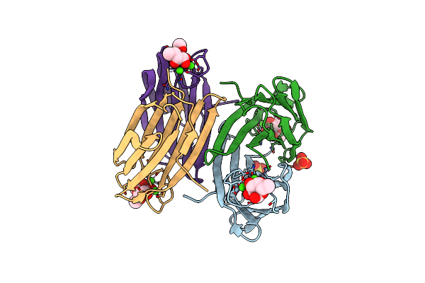
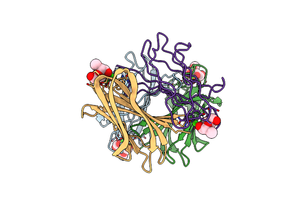
Crystal Structure Of Pa-Il Lectin Complexed With Agal13Bgal14Glc At 1.9 A Resolution
Organism: Pseudomonas aeruginosa
Method: X-RAY DIFFRACTION Resolution:1.90 Å Release Date: 2008-09-16 Classification: SUGAR BINDING PROTEIN Ligands: CA, EDO, BGC |
 |
Mutant (S22A) Of Pseudomonas Aeruginosa Lectin Ii (Pa-Iil) Complexed With Methyl-A-L-Fucopyranoside
Organism: Pseudomonas aeruginosa
Method: X-RAY DIFFRACTION Resolution:1.70 Å Release Date: 2007-06-12 Classification: LECTIN Ligands: CA, SO4, FUC, MFU |
 |
Mutant (S22A) Of Pseudomonas Aeruginosa Lectin Ii (Pa-Iil) Complexed With Methyl-A-L-Mannopyranoside
Organism: Pseudomonas aeruginosa
Method: X-RAY DIFFRACTION Resolution:1.30 Å Release Date: 2007-06-12 Classification: LECTIN Ligands: CA, SO4, FUC, MMA |
 |
Mutant (S23A) Of Pseudomonas Aeruginosa Lectin Ii (Pa-Iil) Complexed With Methyl-A-L-Fucopyranoside
Organism: Pseudomonas aeruginosa
Method: X-RAY DIFFRACTION Resolution:1.30 Å Release Date: 2007-06-12 Classification: LECTIN Ligands: MFU, CA |
 |
Mutant (G24N) Of Pseudomonas Aeruginosa Lectin Ii (Pa-Iil) Complexed With Methyl-A-L-Fucopyranoside
Organism: Pseudomonas aeruginosa
Method: X-RAY DIFFRACTION Resolution:1.50 Å Release Date: 2007-06-12 Classification: LECTIN Ligands: GOL, SO4, CA, MFU |
 |
Mutant (G24N) Of Pseudomonas Aeruginosa Lectin Ii (Pa-Iil) Complexed With Methyl-B-D-Mannoyranoside
Organism: Pseudomonas aeruginosa
Method: X-RAY DIFFRACTION Resolution:1.70 Å Release Date: 2007-06-12 Classification: LECTIN Ligands: MMA, CA |
 |
1.1A Structure Of Chromobacterium Violaceum Lectin Cv2L In Complex With Alpha-Methyl-Fucoside
Organism: Chromobacterium violaceum
Method: X-RAY DIFFRACTION Resolution:1.10 Å Release Date: 2006-05-25 Classification: LECTIN Ligands: CA, MFU |
 |
1.0A Structure Of Chromobacterium Violaceum Lectin In Complex With Alpha-Methyl-Mannoside
Organism: Chromobacterium violaceum
Method: X-RAY DIFFRACTION Resolution:1.00 Å Release Date: 2006-05-25 Classification: LECTIN Ligands: CA, MMA |
 |
Pseudomonas Aeruginosa Lectin Ii (Pa-Iil)Complexed With Lacto-N-Neo- Fucopentaose V(Lnpfv)
Organism: Pseudomonas aeruginosa
Method: X-RAY DIFFRACTION Resolution:1.05 Å Release Date: 2005-03-31 Classification: SUGAR BINDING PROTEIN Ligands: SO4, CA, GOL |
 |
Structure Of Pseudomonas Aeruginosa Lectin Ii (Pa-Iil)Complexed With Lewisa Trisaccharide
Organism: Pseudomonas aeruginosa
Method: X-RAY DIFFRACTION Resolution:1.75 Å Release Date: 2005-03-31 Classification: SUGAR BINDING PROTEIN Ligands: CA, SO4, GOL |
 |
High Affinity Fucose Binding Of Pseudomonas Aeruginosa Lectin Ii: 1.0 A Crystal Structure Of The Complex
Organism: Pseudomonas aeruginosa
Method: X-RAY DIFFRACTION Resolution:1.00 Å Release Date: 2004-12-08 Classification: LECTIN Ligands: SO4, CA, FUC |