
Search Count: 12
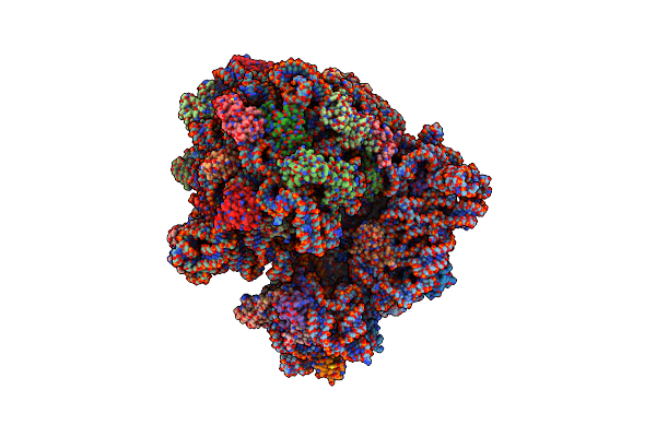 |
Cryo-Em Structure Of The E.Coli 70S Ribosome In Complex With The Antibiotic Myxovalargin B.
Organism: Myxococcus fulvus, Escherichia coli k-12
Method: ELECTRON MICROSCOPY Release Date: 2023-01-25 Classification: RIBOSOME Ligands: MG, ZN, FME, SPD |
 |
Cryo-Em Structure Of The E.Coli 50S Ribosomal Subunit In Complex With The Antibiotic Myxovalargin A.
Organism: Myxococcus fulvus, Escherichia coli k-12
Method: ELECTRON MICROSCOPY Release Date: 2023-01-18 Classification: RIBOSOME Ligands: MG, ZN |
 |
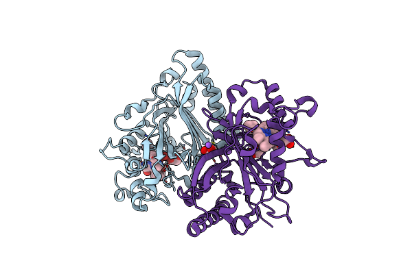
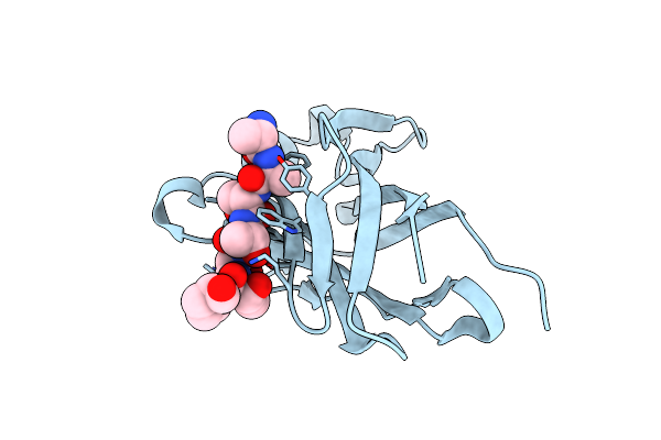
X-Ray Structure Of Trypanosoma Cruzi Pex14 In Complex With A Pex5-Pex14 Ppi Inhibitor
Organism: Trypanosoma cruzi
Method: X-RAY DIFFRACTION Resolution:2.18 Å Release Date: 2022-11-23 Classification: SIGNALING PROTEIN Ligands: GOL, ET7 |
 |
Organism: Severe acute respiratory syndrome coronavirus 2
Method: X-RAY DIFFRACTION Resolution:2.68 Å Release Date: 2022-02-02 Classification: HYDROLASE Ligands: PRL, EDO, ZN, SO4 |
 |
Crystal Structure Of Dyp-Type Peroxidase From Dictyostelium Discoideum In Complex With An Activated Form Of Oxygen
Organism: Dictyostelium discoideum
Method: X-RAY DIFFRACTION Resolution:1.70 Å Release Date: 2021-07-14 Classification: HYDROLASE Ligands: HEM, EDO, NA, OXY |
 |
Dictyostelium Discoideum Dye Decolorizing Peroxidase Dypa In Complex With Cyanide.
Organism: Dictyostelium discoideum
Method: X-RAY DIFFRACTION Resolution:1.85 Å Release Date: 2021-07-14 Classification: HYDROLASE Ligands: HEM, EDO, CYN |
 |
Dictyostelium Discoideum Dye Decolorizing Peroxidase Dypa In Complex With An Activated Form Of Oxygen And Veratryl Alcohol
Organism: Dictyostelium discoideum
Method: X-RAY DIFFRACTION Resolution:1.60 Å Release Date: 2021-07-14 Classification: HYDROLASE Ligands: HEM, VOH, EDO, OXY |
 |
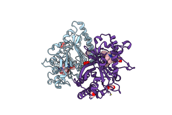
High Resolution Crystal Structure Of N-Terminal Domain Of Pex14 From Trypanosoma Brucei In Complex With The Fist Compound With Sub-Micromolar Trypanocidal Activity
Organism: Trypanosoma brucei brucei
Method: X-RAY DIFFRACTION Resolution:1.20 Å Release Date: 2020-01-01 Classification: SIGNALING PROTEIN Ligands: FTW, BME, CL |
 |
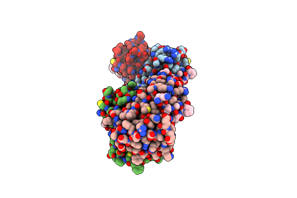
Co-Crystal Structure Of Human Spop Math Domain (Wild-Type) And Human Brd3 Fragment
Organism: Homo sapiens
Method: X-RAY DIFFRACTION Resolution:1.90 Å Release Date: 2019-05-08 Classification: LIGASE |
 |
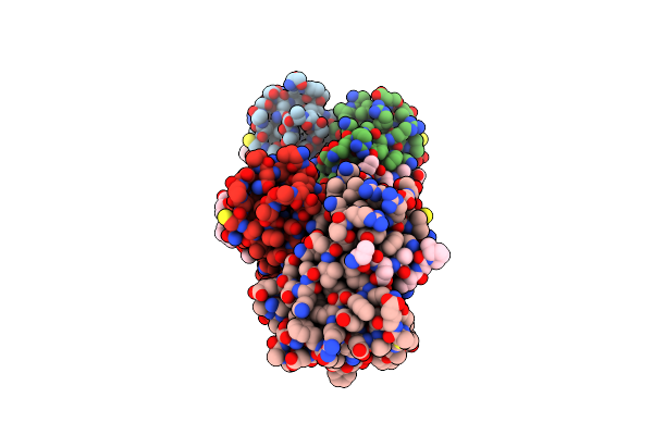
Co-Crystal Structure Of Human Spop Math Domain (E47K) And Human Brd3 Fragment
Organism: Homo sapiens
Method: X-RAY DIFFRACTION Resolution:1.81 Å Release Date: 2019-05-08 Classification: LIGASE |
 |
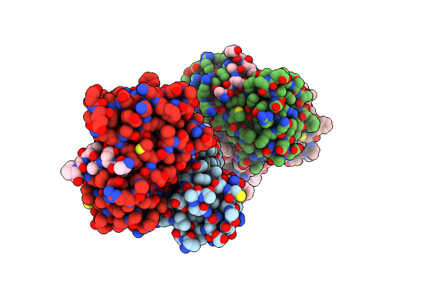
Co-Crystal Structure Of Human Spop Math Domain (M117V) And Human Brd3 Fragment
Organism: Homo sapiens
Method: X-RAY DIFFRACTION Resolution:1.85 Å Release Date: 2019-05-08 Classification: LIGASE |
 |
Co-Crystal Structure Of Human Spop Math Domain (D140N) And Human Brd3 Fragment
Organism: Homo sapiens
Method: X-RAY DIFFRACTION Resolution:2.20 Å Release Date: 2019-05-08 Classification: LIGASE |