
Search Count: 16
 |
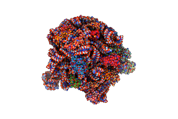
Organism: Homo sapiens, Severe acute respiratory syndrome coronavirus 2
Method: ELECTRON MICROSCOPY Release Date: 2025-04-02 Classification: VIRAL PROTEIN Ligands: NAG |
 |
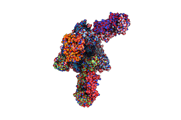
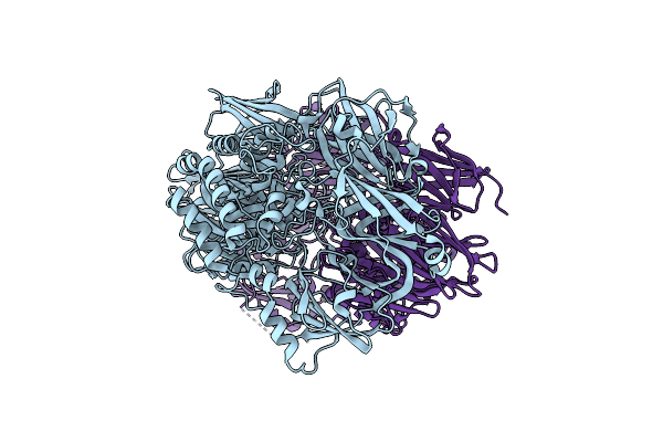
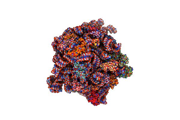
Alpha Sars-Cov-2 Spike Protein Rbd-Down In Complex With Regn10987 Fab Homologue (Local Refinement)
Organism: Severe acute respiratory syndrome coronavirus 2, Homo sapiens
Method: ELECTRON MICROSCOPY Resolution:3.60 Å Release Date: 2025-04-02 Classification: VIRAL PROTEIN |
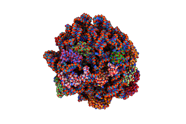 |
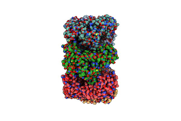
E. Coli 50S Ribosomal Subunit In Complex With Pramp Rumicidin-2 (Focused Refinement)
Organism: Escherichia coli, Beatragus hunteri
Method: ELECTRON MICROSCOPY Release Date: 2025-01-22 Classification: RIBOSOME Ligands: ZN, MG, K, MS6 |
 |
Archaeal Highly Thermostable Gh35 Family Beta-Galactosidase From Desulfurococcus Amyloliticus
Organism: Desulfurococcus amylolyticus
Method: ELECTRON MICROSCOPY Release Date: 2024-10-16 Classification: HYDROLASE |
 |
Organism: Desulfurococcus amylolyticus
Method: X-RAY DIFFRACTION Resolution:2.15 Å Release Date: 2024-08-14 Classification: HYDROLASE |
 |
Inactivated Tick-Borne Encephalitis Virus (Tbev) Vaccine Strain Sofjin-Chumakov
Organism: Orthoflavivirus encephalitidis
Method: ELECTRON MICROSCOPY Release Date: 2024-06-05 Classification: VIRAL PROTEIN Ligands: NAG |
 |
Organism: Tick-borne encephalitis virus (strain sofjin)
Method: ELECTRON MICROSCOPY Release Date: 2024-05-22 Classification: VIRUS Ligands: NAG |
 |
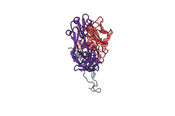
Organism: Severe acute respiratory syndrome coronavirus 2, Homo sapiens
Method: ELECTRON MICROSCOPY Release Date: 2023-04-19 Classification: VIRAL PROTEIN |
 |
Hepatitis B Virus Core Antigen (Hbc) With The Insertion Of Four External Domains Of The Influenza A M2 Protein (Hbc/4M2E) With T=4 Topology
Organism: Hepatitis b virus adw/991, Influenza a virus (a/malaya/302/1954(h1n1)), Hepatitis b virus genotype d subtype ayw (isolate france/tiollais/1979)
Method: ELECTRON MICROSCOPY Release Date: 2022-12-28 Classification: VIRUS LIKE PARTICLE |
 |
Hepatitis B Virus Core Antigen (Hbc) With The Insertion Of Four External Domains Of The Influenza A M2 Protein (Hbc/4M2E) With T=3 Topology
Organism: Hepatitis b virus, Influenza a virus (strain a/malaysia:malaya/302/1954 h1n1)
Method: ELECTRON MICROSCOPY Release Date: 2022-12-28 Classification: VIRUS LIKE PARTICLE |
 |
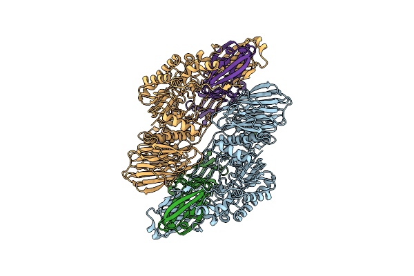
Cryo-Em Structure Of The Groel-Groes Complex With Adp Bound To Both Rings ("Wide" Conformation).
Organism: Escherichia coli (strain k12)
Method: ELECTRON MICROSCOPY Release Date: 2021-11-24 Classification: CHAPERONE Ligands: ADP, MG |
 |
Cryo-Em Structure Of The Groel-Groes Complex With Adp Bound To Both Rings ("Tight" Conformation).
Organism: Escherichia coli (strain k12)
Method: ELECTRON MICROSCOPY Release Date: 2021-11-24 Classification: CHAPERONE Ligands: ADP, MG |
 |
E. Coli 70S Ribosome In Complex With Dirithromycin, And Deacylated Trna(Imet) (Focused Classification).
Organism: Escherichia coli k-12
Method: ELECTRON MICROSCOPY Release Date: 2020-11-04 Classification: RIBOSOME Ligands: DI0 |
 |
E. Coli 70S Ribosome In Complex With Dirithromycin, Fmet-Phe-Trna(Phe) And Deacylated Trna(Imet) (Focused Classification).
Organism: Escherichia coli k-12, Escherichia coli (strain k12), Saccharomyces cerevisiae
Method: ELECTRON MICROSCOPY Release Date: 2020-11-04 Classification: RIBOSOME Ligands: DI0 |
 |
E. Coli 50S Ribosomal Subunit In Complex With Dirithromycin, Fmet-Phe-Trna(Phe) And Deacylated Trna(Imet).
Organism: Escherichia coli (strain k12), Saccharomyces cerevisiae
Method: ELECTRON MICROSCOPY Release Date: 2020-07-22 Classification: RIBOSOME Ligands: MG, DI0 |
 |
Organism: Pseudomonas phage obp
Method: ELECTRON MICROSCOPY Release Date: 2019-08-28 Classification: CHAPERONE |