
Search Count: 19
 |
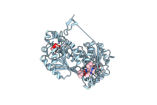
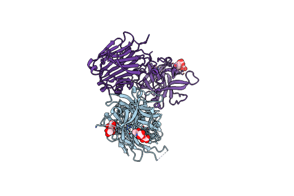
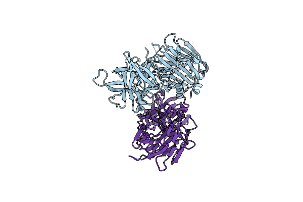
Fusion Construct Of Pqse And Rhlr In Complex With The Synthetic Antagonist Mbtl
Organism: Pseudomonas aeruginosa pao1
Method: X-RAY DIFFRACTION Resolution:3.46 Å Release Date: 2022-12-14 Classification: TRANSCRIPTION Ligands: FE, K5G |
 |
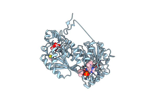
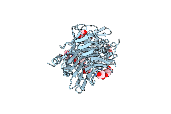
Organism: Pseudomonas aeruginosa pao1
Method: X-RAY DIFFRACTION Resolution:1.65 Å Release Date: 2022-12-14 Classification: HYDROLASE Ligands: FE, CAC, BEZ |
 |
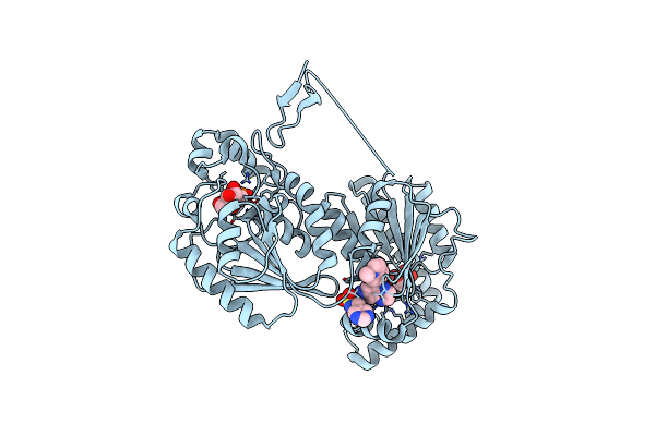
Pross Optimitzed Variant Of Rhlr (75 Mutations) In Complex With The Synthetic Antagonist Mbtl
Organism: Pseudomonas aeruginosa pao1
Method: X-RAY DIFFRACTION Resolution:2.15 Å Release Date: 2022-12-14 Classification: TRANSCRIPTION Ligands: K5G |
 |
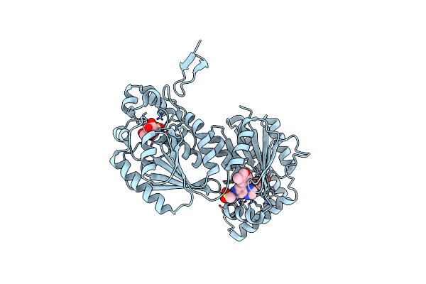
Pross Optimitzed Variant Of Rhlr (75 Mutations) In Complex With Native Autoinducer C4-Hsl
Organism: Pseudomonas aeruginosa pao1
Method: X-RAY DIFFRACTION Resolution:3.49 Å Release Date: 2022-12-14 Classification: TRANSCRIPTION Ligands: HL4 |
 |
Pross Optimitzed Variant Of Rhlr (61 Mutations) In Complex With The Synthetic Antagonist Mbtl
Organism: Pseudomonas aeruginosa pao1
Method: X-RAY DIFFRACTION Resolution:3.10 Å Release Date: 2022-12-14 Classification: TRANSCRIPTION Ligands: K5G |
 |
Organism: Pseudomonas aeruginosa pao1
Method: X-RAY DIFFRACTION Resolution:3.06 Å Release Date: 2022-12-14 Classification: TRANSCRIPTION Ligands: FE, K5G |
 |
Organism: Pseudomonas aeruginosa pao1
Method: X-RAY DIFFRACTION Resolution:3.06 Å Release Date: 2022-12-14 Classification: SIGNALING PROTEIN Ligands: FE, HL4 |
 |
Organism: Homo sapiens
Method: X-RAY DIFFRACTION Resolution:2.11 Å Release Date: 2019-01-23 Classification: TRANSFERASE Ligands: H9Z, POP, FLC, F6P, DMS |
 |
Organism: Homo sapiens
Method: X-RAY DIFFRACTION Resolution:2.51 Å Release Date: 2019-01-23 Classification: TRANSFERASE Ligands: HAT, POP, FLC, F6P, BME, PO4 |
 |
Organism: Homo sapiens
Method: X-RAY DIFFRACTION Resolution:2.44 Å Release Date: 2019-01-23 Classification: TRANSFERASE Ligands: PO4, FLC, HAW, F6P |
 |
Organism: Homo sapiens
Method: X-RAY DIFFRACTION Resolution:2.60 Å Release Date: 2019-01-23 Classification: TRANSFERASE Ligands: HAK, POP, FLC, F6P, DMS |
 |
Organism: Homo sapiens
Method: X-RAY DIFFRACTION Resolution:2.36 Å Release Date: 2018-11-14 Classification: TRANSFERASE Ligands: GV2, POP, FLC, F6P |
 |
Organism: Homo sapiens
Method: X-RAY DIFFRACTION Resolution:1.96 Å Release Date: 2018-11-14 Classification: TRANSFERASE Ligands: GV5, POP, F6P, FLC, DMS |
 |
Organism: Homo sapiens
Method: X-RAY DIFFRACTION Resolution:2.28 Å Release Date: 2018-11-14 Classification: TRANSFERASE Ligands: GV8, F6P, PO4, FLC |
 |
Cell Entry Of Botulinum Neurotoxin Type C Is Dependent Upon Interaction With Two Ganglioside Molecules
Organism: Clostridium botulinum
Method: X-RAY DIFFRACTION Resolution:2.15 Å Release Date: 2011-06-08 Classification: HYDROLASE Ligands: SLB, SIA |
 |
Cell Entry Of Botulinum Neurotoxin Type C Is Dependent Upon Interaction With Two Ganglioside Molecules
Organism: Clostridium botulinum
Method: X-RAY DIFFRACTION Resolution:2.20 Å Release Date: 2011-06-08 Classification: HYDROLASE |
 |
Organism: Clostridium botulinum
Method: X-RAY DIFFRACTION Resolution:1.72 Å Release Date: 2010-09-08 Classification: HYDROLASE Ligands: GOL |
 |
Crystal Structure Of Botulinum Neurotoxin Serotype D Ligand Binding Domain In Complex With N-Acetylneuraminic Acid
Organism: Clostridium botulinum
Method: X-RAY DIFFRACTION Resolution:2.00 Å Release Date: 2010-09-08 Classification: HYDROLASE Ligands: SLB, GOL |
 |
Organism: Clostridium botulinum
Method: X-RAY DIFFRACTION Resolution:1.75 Å Release Date: 2007-09-11 Classification: TOXIN Ligands: ZN |