
Search Count: 32
 |
Organism: Homo sapiens
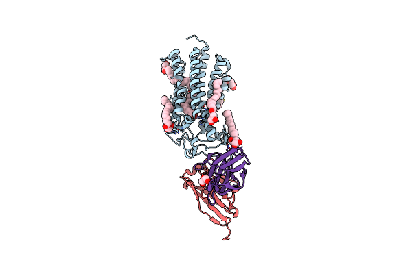
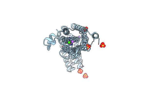
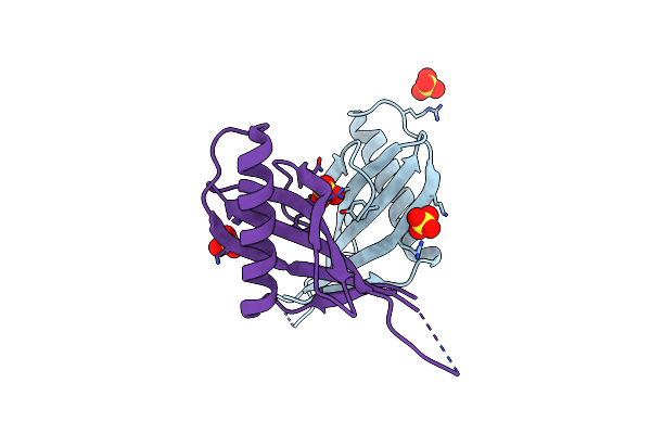
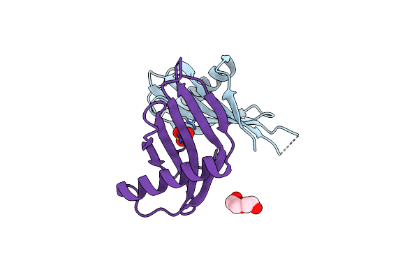
Method: X-RAY DIFFRACTION Release Date: 2025-09-24 Classification: HYDROLASE Ligands: MG, EDO, A1JD4 |
 |
Organism: Homo sapiens
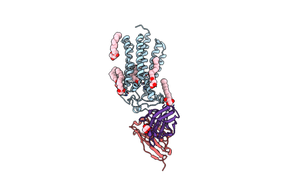
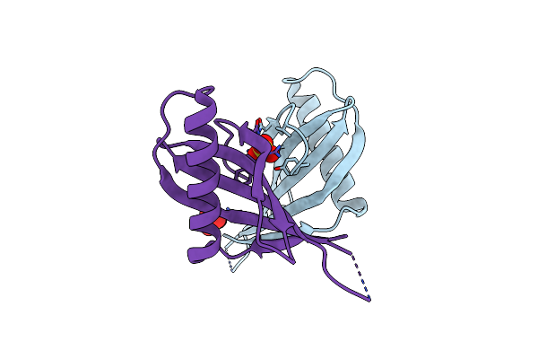
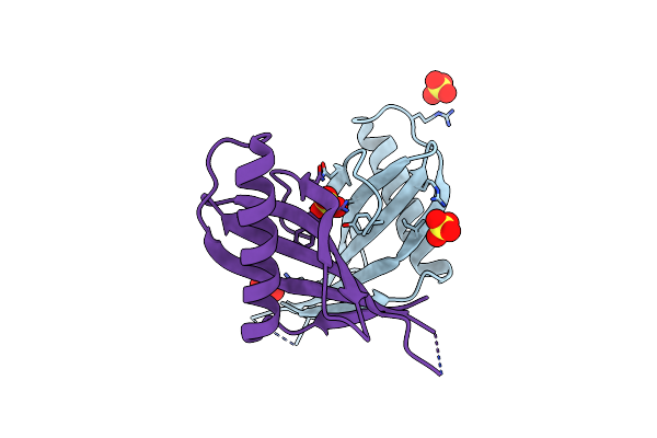
Method: X-RAY DIFFRACTION Release Date: 2025-09-24 Classification: HYDROLASE Ligands: A1JED, TEW, MG |
 |
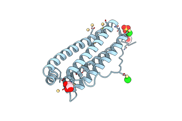
Cryogenic Human Adiponectin Receptor 2 (Adipor2) At 2.4 A Resolution Determined By Serial Crystallography (Ssx) Using Crystaldirect
Organism: Homo sapiens
Method: X-RAY DIFFRACTION Resolution:2.40 Å Release Date: 2021-05-12 Classification: MEMBRANE PROTEIN Ligands: ZN, OLB, OLA, GOL |
 |
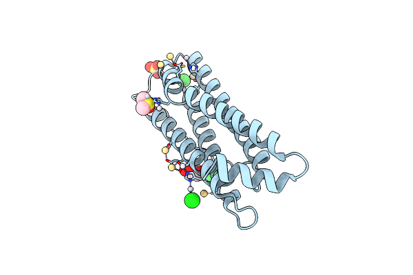
Room Temperature Structure Of Human Adiponectin Receptor 2 (Adipor2) At 2.9 A Resolution Determined By Serial Crystallography (Ssx) Using Crystaldirect
Organism: Homo sapiens
Method: X-RAY DIFFRACTION Resolution:2.90 Å Release Date: 2021-05-12 Classification: MEMBRANE PROTEIN Ligands: ZN, OLB, OLA, GOL |
 |
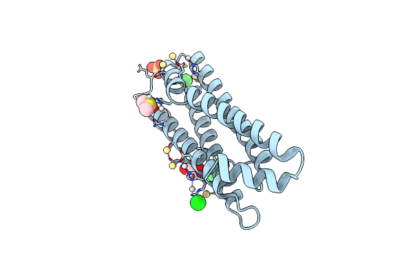
Cryogenic Human Adiponectin Receptor 2 (Adipor2) With Gd-Do3 Ligand Determined By Serial Crystallography (Ssx) Using Crystaldirect
Organism: Homo sapiens
Method: X-RAY DIFFRACTION Resolution:3.03 Å Release Date: 2021-05-12 Classification: MEMBRANE PROTEIN Ligands: ZN, OLA, OLB, GD, DO3 |
 |
Cryogenic Human Adiponectin Receptor 2 (Adipor2) With Tb-Xo4 Ligand Determined By Serial Crystallography (Ssx) Using Crystaldirect
Organism: Homo sapiens
Method: X-RAY DIFFRACTION Resolution:3.01 Å Release Date: 2021-05-12 Classification: MEMBRANE PROTEIN Ligands: ZN, OLA, OLB, 7MT |
 |
Cryogenic Human Alkaline Ceramidase 3 (Acer3) At 2.6 A Resolution Determined By Serial Crystallography (Ssx) Using Crystaldirect
Organism: Homo sapiens
Method: X-RAY DIFFRACTION Resolution:2.60 Å Release Date: 2021-05-12 Classification: MEMBRANE PROTEIN Ligands: ZN, SO4, NA, CA, MG |
 |
Organism: Equus caballus
Method: X-RAY DIFFRACTION Resolution:1.43 Å Release Date: 2019-02-06 Classification: METAL TRANSPORT Ligands: CD, CL, SO4, GOL |
 |
The X-Ray Structure Of The Horse Spleen Ferritin Nanocage Containing Pt, Obtained Upon Encapsulation Of A Pt(Ii) Terpyridine Compound Within The Protein Cage
Organism: Equus caballus
Method: X-RAY DIFFRACTION Resolution:1.33 Å Release Date: 2018-12-19 Classification: METAL TRANSPORT Ligands: CD, CL, PT, SO4, GOL, DMS |
 |
The X-Ray Structure Of The Horse Spleen Ferritin Nanocage Containing Pt, Obtained Upon Encapsulation Of A Pt(Ii) Terpyridine Compound Within The Protein Cage
Organism: Equus caballus
Method: X-RAY DIFFRACTION Resolution:1.58 Å Release Date: 2018-12-19 Classification: METAL TRANSPORT Ligands: CD, CL, PT, SO4, GOL, DMS |
 |
Organism: Dioscoreophyllum cumminsii
Method: X-RAY DIFFRACTION Resolution:2.05 Å Release Date: 2018-01-10 Classification: PLANT PROTEIN Ligands: SO4 |
 |
Organism: Dioscoreophyllum cumminsii
Method: X-RAY DIFFRACTION Resolution:2.60 Å Release Date: 2018-01-10 Classification: PLANT PROTEIN Ligands: SO4 |
 |
Organism: Dioscoreophyllum cumminsii
Method: X-RAY DIFFRACTION Resolution:1.72 Å Release Date: 2018-01-10 Classification: PLANT PROTEIN Ligands: SO4, PEG |
 |
Organism: Dioscoreophyllum cumminsii
Method: X-RAY DIFFRACTION Resolution:2.06 Å Release Date: 2018-01-10 Classification: PLANT PROTEIN Ligands: SO4 |
 |
Organism: Dioscoreophyllum cumminsii
Method: X-RAY DIFFRACTION Resolution:2.02 Å Release Date: 2018-01-10 Classification: PLANT PROTEIN Ligands: SO4, PEG |
 |
Organism: Homo sapiens
Method: X-RAY DIFFRACTION Resolution:2.95 Å Release Date: 2016-11-30 Classification: protein/dna Ligands: NAG, 0G6, NA |
 |
Organism: Homo sapiens
Method: X-RAY DIFFRACTION Resolution:3.59 Å Release Date: 2016-11-30 Classification: protein/dna Ligands: NAG, 0G6, NA |
 |
Organism: Dioscoreophyllum cumminsii
Method: X-RAY DIFFRACTION Resolution:1.70 Å Release Date: 2016-10-05 Classification: PLANT PROTEIN Ligands: SO4 |
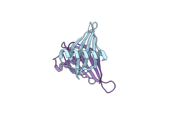 |
Crystal Structure Of A Single Chain Monellin Mutant: E23Q/Q28K/C41S/Y65R-Mnei
Organism: Dioscoreophyllum cumminsii
Method: X-RAY DIFFRACTION Resolution:1.55 Å Release Date: 2016-10-05 Classification: PLANT PROTEIN |
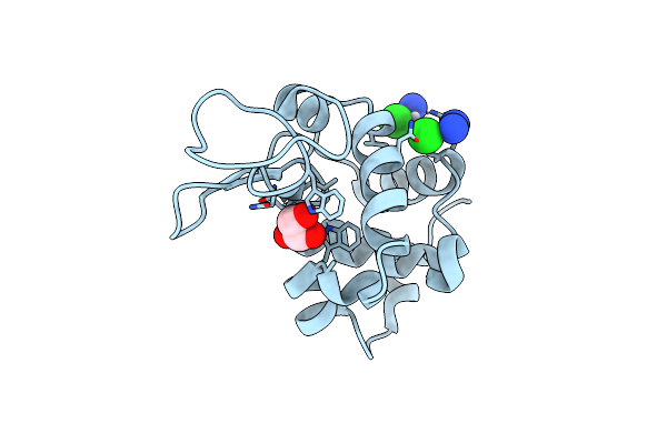 |
X-Ray Structure Of The Adduct Between Hen Egg White Lysozyme And Cisplatin Upon 24 Hours Of Incubation At 20 Degrees
Organism: Gallus gallus
Method: X-RAY DIFFRACTION Resolution:1.85 Å Release Date: 2016-04-13 Classification: HYDROLASE Ligands: GOL, CPT |