
Search Count: 17
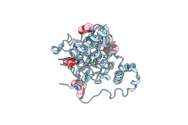 |
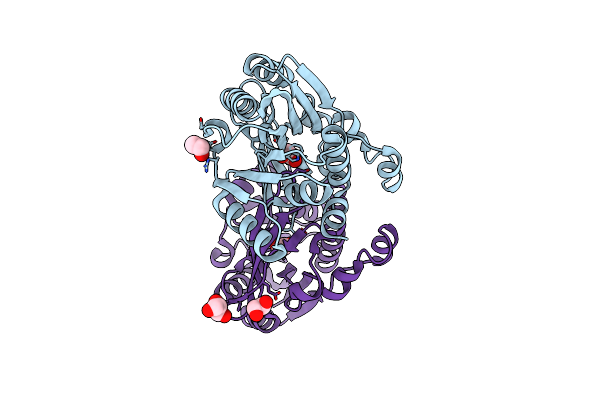
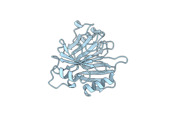
Crystal Structure Of Burkholderia Glumae Toxoflavin Biosynthesis Protein Toxd
Organism: Burkholderia glumae
Method: X-RAY DIFFRACTION Resolution:1.80 Å Release Date: 2023-05-10 Classification: UNKNOWN FUNCTION Ligands: CIT, CA, PG4, 1PE |
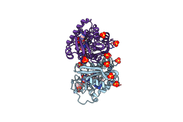 |
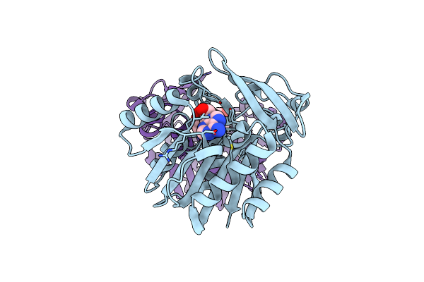
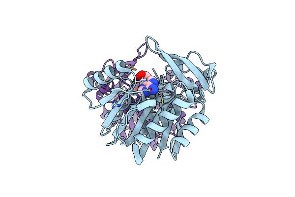
Crystal Structure Of Burkholderia Thailandensis 1,6-Didemethyltoxoflavin-N1-Methyltransferase With Bound 1,6-Didemethyltoxoflavin And S-Adenosylhomocysteine
Organism: Burkholderia thailandensis (strain atcc 700388 / dsm 13276 / cip 106301 / e264)
Method: X-RAY DIFFRACTION Resolution:1.77 Å Release Date: 2017-12-13 Classification: TRANSFERASE Ligands: SAH, AZ8, SO4, EPE |
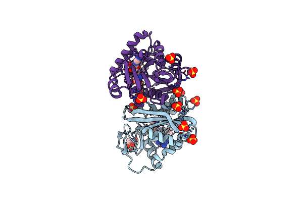 |
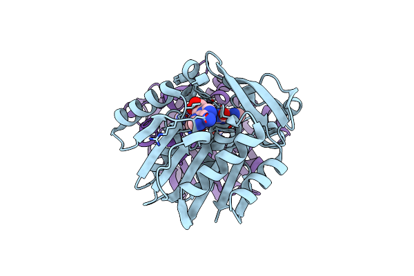
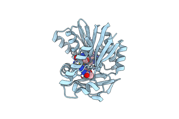
Crystal Structure Of Burkholderia Thailandensis 1,6-Didemethyltoxoflavin-N1-Methyltransferase With Bound S-Adenosylhomocysteine
Organism: Burkholderia thailandensis (strain atcc 700388 / dsm 13276 / cip 106301 / e264)
Method: X-RAY DIFFRACTION Resolution:1.39 Å Release Date: 2017-12-13 Classification: TRANSFERASE Ligands: SAH, SO4, EPE |
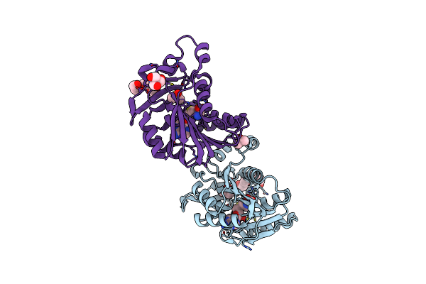 |
Crystal Structure Of Burkholderia Glumae Toxa Y7F Mutant With Bound S-Adenosylhomocysteine (Sah)
Organism: Burkholderia glumae
Method: X-RAY DIFFRACTION Resolution:1.52 Å Release Date: 2016-05-11 Classification: TRANSFERASE Ligands: SAH, DMS, PG4, TRS |
 |
Organism: Burkholderia glumae
Method: X-RAY DIFFRACTION Resolution:1.57 Å Release Date: 2016-05-11 Classification: TRANSFERASE Ligands: EDO, GOL |
 |
Crystal Structure Of Burkholderia Glumae Toxa Y7F Mutant With Bound S-Adenosylhomocysteine (Sah) And Toxoflavin
Organism: Burkholderia glumae
Method: X-RAY DIFFRACTION Resolution:1.77 Å Release Date: 2016-05-04 Classification: TRANSFERASE Ligands: SAH, TOF, TRS |
 |
Crystal Structure Of Burkholderia Glumae Toxa With Bound S-Adenosylhomocysteine (Sah)
Organism: Burkholderia glumae
Method: X-RAY DIFFRACTION Resolution:1.60 Å Release Date: 2016-05-04 Classification: TRANSFERASE Ligands: SAH |
 |
Crystal Structure Of Burkholderia Glumae Toxa With Bound S-Adenosylhomocysteine (Sah) And 1,6-Didemethyltoxoflavin
Organism: Burkholderia glumae
Method: X-RAY DIFFRACTION Resolution:1.55 Å Release Date: 2016-05-04 Classification: TRANSFERASE Ligands: SAH, AZ8 |
 |
Crystal Structure Of Burkholderia Glumae Toxa With Bound S-Adenosylhomocysteine (Sah) And Toxoflavin
Organism: Burkholderia glumae
Method: X-RAY DIFFRACTION Resolution:1.95 Å Release Date: 2016-05-04 Classification: TRANSFERASE Ligands: SAH, TOF |
 |
Crystal Structure Of Burkholderia Glumae Toxa Y7A Mutant With Bound S-Adenosylhomocysteine (Sah)
Organism: Burkholderia glumae
Method: X-RAY DIFFRACTION Resolution:1.79 Å Release Date: 2016-05-04 Classification: TRANSFERASE Ligands: SAH |
 |
Crystal Structure Of Burkholderia Glumae Toxa Y7A Mutant With Bound S-Adenosylhomocysteine (Sah)
Organism: Burkholderia glumae
Method: X-RAY DIFFRACTION Resolution:1.93 Å Release Date: 2016-05-04 Classification: TRANSFERASE Ligands: SAH |
 |
Crystal Structure Of Burkholderia Glumae Toxa With Bound S-Adenosylhomocysteine (Sah) And 1-Demethyltoxoflavin
Organism: Burkholderia glumae
Method: X-RAY DIFFRACTION Resolution:1.56 Å Release Date: 2016-05-04 Classification: TRANSFERASE Ligands: SAH, AZ9 |
 |
Organism: Candida albicans
Method: X-RAY DIFFRACTION Resolution:1.60 Å Release Date: 2012-09-19 Classification: TRANSFERASE Ligands: CIT |
 |
Organism: Candida albicans
Method: X-RAY DIFFRACTION Resolution:2.20 Å Release Date: 2012-09-19 Classification: TRANSFERASE |
 |
Organism: Paenibacillus polymyxa
Method: X-RAY DIFFRACTION Resolution:1.34 Å Release Date: 2011-01-12 Classification: LYASE |
 |
Organism: Paenibacillus polymyxa
Method: X-RAY DIFFRACTION Resolution:1.80 Å Release Date: 2011-01-12 Classification: LYASE Ligands: MN |
 |
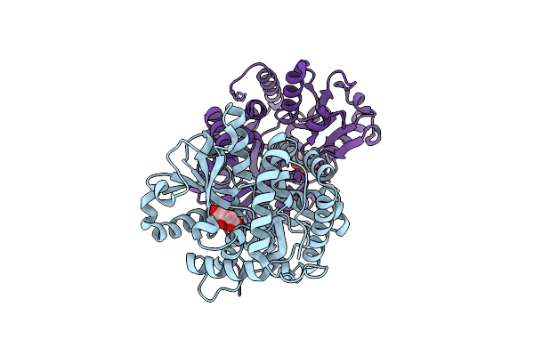
Crystal Structure Of Toxoflavin Lyase (Tfla) Bound To Mn(Ii) And Toxoflavin
Organism: Paenibacillus polymyxa
Method: X-RAY DIFFRACTION Resolution:1.50 Å Release Date: 2011-01-12 Classification: LYASE Ligands: MN, TOF |