
Search Count: 17
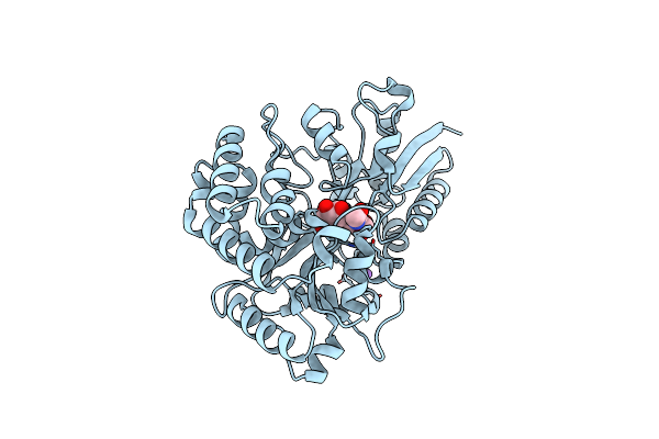 |
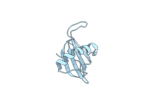
Fucosylated Lacto-N-Biose Binding Protein From Bifidobacterium Longum Subsp. Infantis In Complex With Galacto-N-Biose
Organism: Bifidobacterium longum subsp. infantis
Method: X-RAY DIFFRACTION Release Date: 2025-07-02 Classification: TRANSPORT PROTEIN |
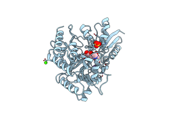 |
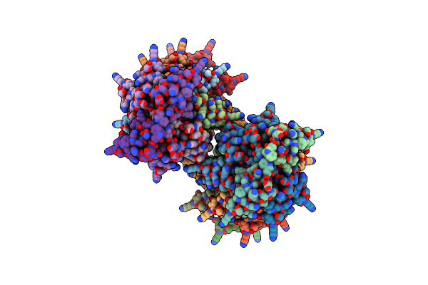
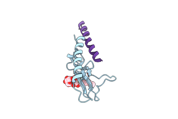
Fucosylated Lacto-N-Biose Binding Protein From Bifidobacterium Longum Subsp. Infantis In Complex With Lacto-N-Biose
Organism: Bifidobacterium longum subsp. infantis
Method: X-RAY DIFFRACTION Release Date: 2025-07-02 Classification: TRANSPORT PROTEIN Ligands: CA, SO4, NA |
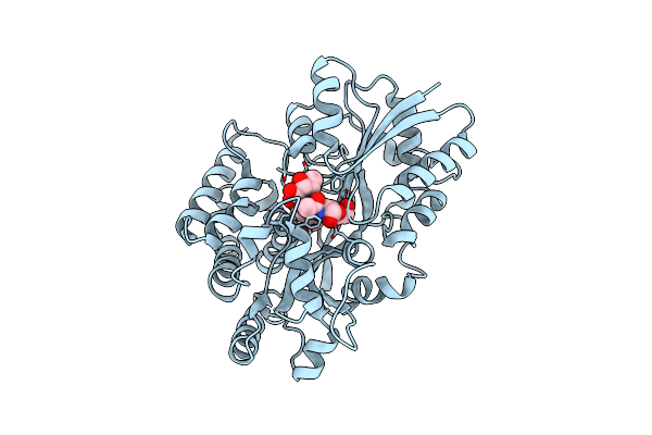 |
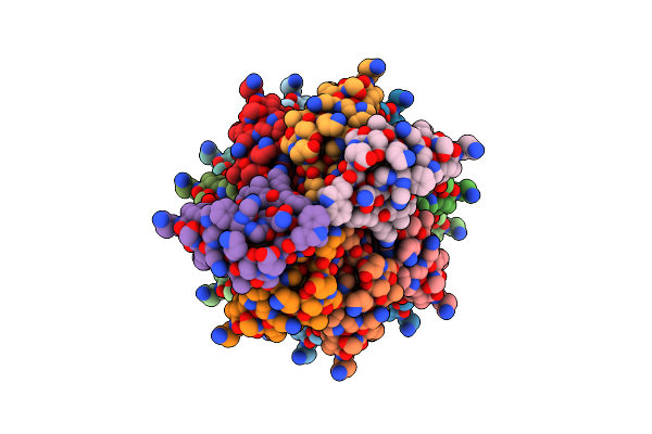
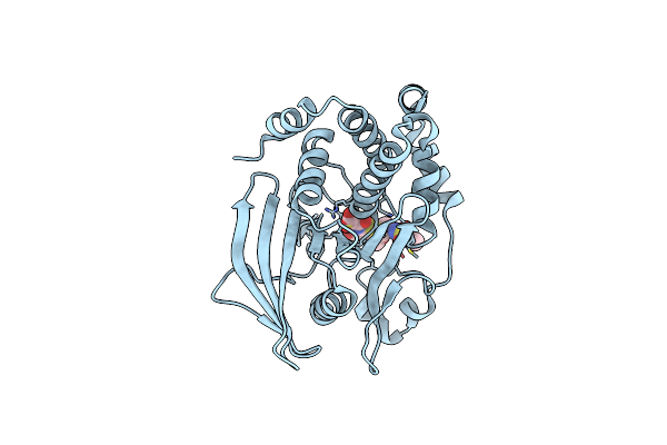
Fucosylated Lacto-N-Biose Binding Protein From Bifidobacterium Longum Subsp. Infantis In Complex With H1 Trisaccharide
Organism: Bifidobacterium longum subsp. infantis
Method: X-RAY DIFFRACTION Release Date: 2025-07-02 Classification: TRANSPORT PROTEIN |
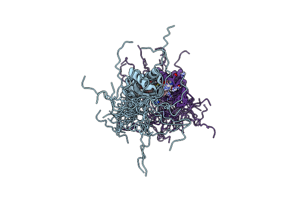 |
Organism: Homo sapiens
Method: SOLUTION NMR Release Date: 2023-05-03 Classification: OXIDOREDUCTASE Ligands: PHE |
 |
Organism: Pseudoplectania nigrella
Method: X-RAY DIFFRACTION Resolution:1.13 Å Release Date: 2022-06-29 Classification: ANTIMICROBIAL PROTEIN Ligands: PEG |
 |
Organism: Pseudoplectania nigrella
Method: ELECTRON MICROSCOPY Release Date: 2022-04-27 Classification: ANTIMICROBIAL PROTEIN |
 |
Organism: Pseudoplectania nigrella
Method: ELECTRON MICROSCOPY Release Date: 2022-04-27 Classification: ANTIMICROBIAL PROTEIN |
 |
Glycoside Hydrolase Family 109 From Akkermansia Muciniphila In Complex With Galnac And Nad+.
Organism: Akkermansia muciniphila
Method: X-RAY DIFFRACTION Resolution:2.13 Å Release Date: 2020-02-26 Classification: HYDROLASE Ligands: A2G, NAD, PGE, 1PE |
 |
Structural Characterization Of A Mannuronic Acid Specific Polysaccharide Family 6 Lyase Enzyme From Human Gut Microbiota
Organism: Bacteroides cellulosilyticus
Method: X-RAY DIFFRACTION Resolution:1.29 Å Release Date: 2019-09-25 Classification: LYASE Ligands: CA, ACT |
 |
Organism: Synthetic construct
Method: X-RAY DIFFRACTION Resolution:2.50 Å Release Date: 2019-07-03 Classification: DE NOVO PROTEIN |
 |
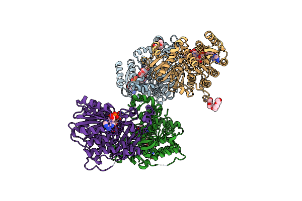
Crystal Structure Of Glucagon-Like Peptide-1 In Complex With The Extracellular Domain Of The Glucagon-Like Peptide-1 Receptor
Organism: Homo sapiens
Method: X-RAY DIFFRACTION Resolution:2.10 Å Release Date: 2009-10-27 Classification: SIGNALING PROTEIN/SIGNALING PROTEIN Ligands: 10M |
 |
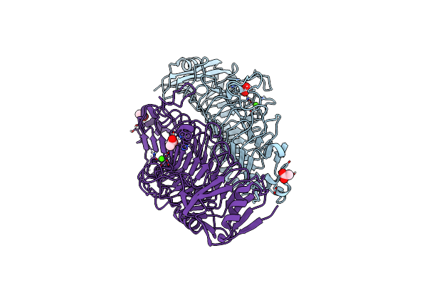
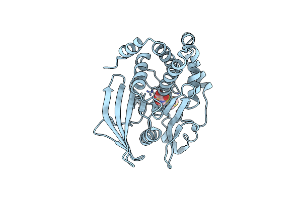
Crystal Structure Of Ptp1B Complexed With 7-(1,1-Dioxo-1H-Benzo[D]Isothiazol-3-Yloxymethyl)-2-(Oxalyl-Amino)-4,7-Dihydro-5H-Thieno[2,3-C]Pyran-3-Carboxylic Acid
Organism: Homo sapiens
Method: X-RAY DIFFRACTION Resolution:2.50 Å Release Date: 2002-05-08 Classification: HYDROLASE Ligands: DBD |
 |
Organism: Homo sapiens
Method: X-RAY DIFFRACTION Resolution:2.56 Å Release Date: 2002-05-08 Classification: HYDROLASE |
 |
Residue 259 Is A Key Determinant Of Substrate Specificity Of Protein-Tyrosine Phosphatase 1B And Alpha
Organism: Homo sapiens
Method: X-RAY DIFFRACTION Resolution:2.13 Å Release Date: 2000-07-04 Classification: HYDROLASE Ligands: COL |
 |
Crystal Structure Of Protein Tyrosine Phosphatase 1B (R47V,D48N) Complexed With 2-(Oxalyl-Amino-4,7-Dihydro-5H-Thieno[2,3-C]Pyran-3-Carboxylic Acid
Organism: Homo sapiens
Method: X-RAY DIFFRACTION Resolution:2.30 Å Release Date: 2000-05-03 Classification: HYDROLASE Ligands: OPA |
 |
Crystal Structure Of Protein Tyrosine Phosphatase 1B Complexed With 2-(Oxalyl-Amino-4,7-Dihydro-5H-Thieno[2,3-C]Pyran-3-Carboxylic Acid
Organism: Homo sapiens
Method: X-RAY DIFFRACTION Resolution:2.10 Å Release Date: 2000-05-03 Classification: HYDROLASE Ligands: OPA |
 |
Crystal Structure Of Protein Tyrosine Phosphatase 1B Complexed With 2-(Oxalyl-Amino)-4,5,6,7-Tetrahydro-Thieno[2,3-C]Pyridine-3-Carboxylic Acid
Organism: Homo sapiens
Method: X-RAY DIFFRACTION Resolution:1.80 Å Release Date: 2000-05-03 Classification: HYDROLASE Ligands: OTA |