
Search Count: 17
 |
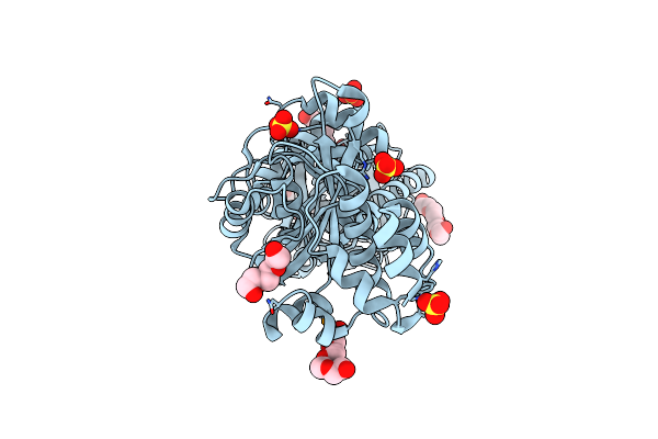
Organism: Haemophilus influenzae
Method: X-RAY DIFFRACTION Resolution:1.90 Å Release Date: 2023-12-27 Classification: SUGAR BINDING PROTEIN Ligands: SLB, ZN |
 |
Organism: Strongylocentrotus purpuratus
Method: ELECTRON MICROSCOPY Release Date: 2023-11-08 Classification: MEMBRANE PROTEIN |
 |
Organism: Strongylocentrotus purpuratus
Method: ELECTRON MICROSCOPY Release Date: 2023-11-08 Classification: MEMBRANE PROTEIN |
 |
Organism: Strongylocentrotus purpuratus
Method: ELECTRON MICROSCOPY Release Date: 2023-11-08 Classification: MEMBRANE PROTEIN |
 |
Organism: Strongylocentrotus purpuratus
Method: ELECTRON MICROSCOPY Release Date: 2023-11-08 Classification: MEMBRANE PROTEIN |
 |
Organism: Strongylocentrotus purpuratus
Method: ELECTRON MICROSCOPY Release Date: 2023-11-08 Classification: MEMBRANE PROTEIN |
 |
Organism: Strongylocentrotus purpuratus
Method: ELECTRON MICROSCOPY Release Date: 2023-11-08 Classification: MEMBRANE PROTEIN Ligands: CMP |
 |
Organism: Strongylocentrotus purpuratus
Method: ELECTRON MICROSCOPY Release Date: 2023-11-08 Classification: MEMBRANE PROTEIN Ligands: CMP |
 |
Organism: Strongylocentrotus purpuratus
Method: ELECTRON MICROSCOPY Release Date: 2023-11-08 Classification: MEMBRANE PROTEIN Ligands: PCG |
 |
Organism: Strongylocentrotus purpuratus
Method: ELECTRON MICROSCOPY Release Date: 2023-11-08 Classification: MEMBRANE PROTEIN Ligands: PCG |
 |
Organism: Sulfurihydrogenibium sp. yo3aop1
Method: X-RAY DIFFRACTION Resolution:2.10 Å Release Date: 2022-11-16 Classification: PEPTIDE BINDING PROTEIN Ligands: SO4, PGE, PG4 |
 |
Organism: Sulfurihydrogenibium
Method: X-RAY DIFFRACTION Resolution:2.20 Å Release Date: 2022-11-16 Classification: HYDROLASE Ligands: PGE, SO4 |
 |
Organism: Sulfurihydrogenibium
Method: X-RAY DIFFRACTION Resolution:3.29 Å Release Date: 2022-11-16 Classification: HYDROLASE Ligands: SO4, GOL |
 |
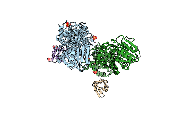
Structure Of The Membrane Domains Of The Sialic Acid Trap Transporter Hisiaqm From Haemophilus Influenzae
Organism: Haemophilus influenzae, Vicugna pacos
Method: ELECTRON MICROSCOPY Release Date: 2022-07-27 Classification: TRANSPORT PROTEIN |
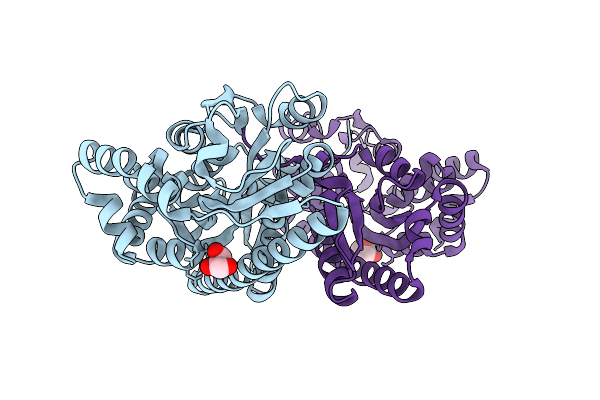 |
Crystal Structure Of Spin Labelled Vcsiap R125A Bound To An Artificial Peptide Ligand.
Organism: Vibrio cholerae serotype o1 (strain atcc 39315 / el tor inaba n16961)
Method: X-RAY DIFFRACTION Resolution:2.20 Å Release Date: 2020-12-30 Classification: TRANSPORT PROTEIN Ligands: GOL |
 |
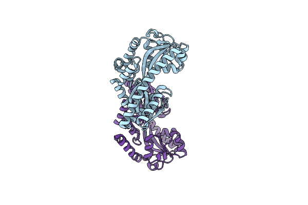
Organism: Vibrio cholerae
Method: X-RAY DIFFRACTION Resolution:1.68 Å Release Date: 2020-12-30 Classification: TRANSPORT PROTEIN Ligands: SLB, GOL, PGE, BGC |
 |
Organism: Vibrio cholerae
Method: X-RAY DIFFRACTION Resolution:2.10 Å Release Date: 2017-01-25 Classification: MEMBRANE PROTEIN |