
Search Count: 117
 |
Organism: Vicugna pacos
Method: X-RAY DIFFRACTION Release Date: 2025-08-27 Classification: IMMUNE SYSTEM Ligands: SO4, CL, GOL, 1PE |
 |
Organism: Vicugna pacos
Method: X-RAY DIFFRACTION Release Date: 2025-08-27 Classification: IMMUNE SYSTEM Ligands: SO4, NA, GOL |
 |
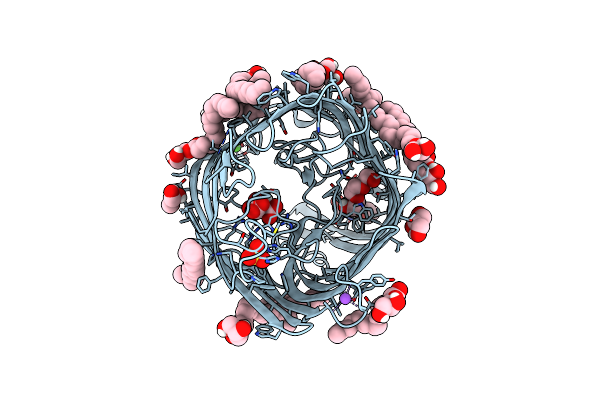
High-Resolution Structure Of The Siderophore Periplasmic Binding Protein Ftsb From Streptococcus Pyogenes
Organism: Streptococcus pyogenes ssi-1
Method: X-RAY DIFFRACTION Resolution:1.11 Å Release Date: 2024-10-09 Classification: METAL BINDING PROTEIN Ligands: P33, PEG, EDO, P6G, ZN, NA, CL |
 |
Structure Of The Siderophore Periplasmic Binding Protein Ftsb From Streptococcus Pyogenes With Ferrichrome Bound
Organism: Streptococcus pyogenes ssi-1
Method: X-RAY DIFFRACTION Resolution:1.80 Å Release Date: 2024-10-09 Classification: METAL BINDING PROTEIN Ligands: FCE |
 |
Structure Of The Siderophore Periplasmic Binding Protein Ftsb From Streptococcus Pyogenes With Bisucaberin Bound
Organism: Streptococcus pyogenes ssi-1
Method: X-RAY DIFFRACTION Resolution:2.00 Å Release Date: 2024-10-09 Classification: METAL BINDING PROTEIN Ligands: OX8, FE |
 |
High-Resolution Structure Of The Siderophore Periplasmic Binding Protein Ftsb From Streptococcus Pyogenes With Ferrioxamine E Bound
Organism: Streptococcus pyogenes ssi-1
Method: X-RAY DIFFRACTION Resolution:1.12 Å Release Date: 2024-10-09 Classification: METAL BINDING PROTEIN Ligands: 6L0, FE, ZN, NA, GOL, EDO |
 |
High-Resolution Structure Of The Siderophore Periplasmic Binding Protein Ftsb From Streptococcus Pyogenes With Ferrioxamine B
Organism: Streptococcus pyogenes ssi-1
Method: X-RAY DIFFRACTION Resolution:1.15 Å Release Date: 2024-10-09 Classification: METAL BINDING PROTEIN Ligands: 0UE, ZN, EDO, 03S, NA |
 |
High-Resolution Structure Of The Siderophore Periplasmic Binding Protein Ftsb Mutant Y137A From Streptococcus Pyogenes
Organism: Streptococcus pyogenes ssi-1
Method: X-RAY DIFFRACTION Resolution:1.15 Å Release Date: 2024-10-09 Classification: METAL BINDING PROTEIN Ligands: P33, PEG, ZN, SO4, CL, NA |
 |
Structure Of The Siderophore Periplasmic Binding Protein Ftsb Mutant Y137A From Streptococcus Pyogenes With Ferrioxamine E Bound
Organism: Streptococcus pyogenes ssi-1
Method: X-RAY DIFFRACTION Resolution:1.85 Å Release Date: 2024-10-09 Classification: METAL BINDING PROTEIN Ligands: 6L0, FE, GOL |
 |
High-Resolution Structure Of The Siderophore Periplasmic Binding Protein Ftsb From Streptococcus Pyogenes With Ferrioxamine E Bound (Crystal Form 2)
Organism: Streptococcus pyogenes ssi-1
Method: X-RAY DIFFRACTION Resolution:1.95 Å Release Date: 2024-10-09 Classification: METAL BINDING PROTEIN Ligands: 6L0, FE, CL |
 |
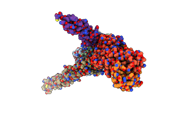
Organism: Homo sapiens, Severe acute respiratory syndrome coronavirus 2
Method: X-RAY DIFFRACTION Resolution:3.30 Å Release Date: 2024-05-15 Classification: VIRAL PROTEIN/IMMUNE SYSTEM |
 |
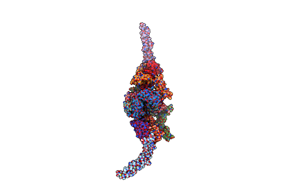
Organism: Homo sapiens
Method: X-RAY DIFFRACTION Resolution:2.70 Å Release Date: 2024-05-08 Classification: IMMUNE SYSTEM |
 |
Organism: Homo sapiens
Method: X-RAY DIFFRACTION Resolution:2.80 Å Release Date: 2024-05-08 Classification: IMMUNE SYSTEM Ligands: GOL |
 |
In Meso Structure Of The Alginate Exporter, Alge, From Pseudomonas Aeruginosa In 7.10 Monoacylglycerol
Organism: Pseudomonas aeruginosa
Method: X-RAY DIFFRACTION Resolution:1.45 Å Release Date: 2024-04-03 Classification: MEMBRANE PROTEIN Ligands: CIT, P6G, A1H2K, GOL, A1H52, NA, SO4, CA |
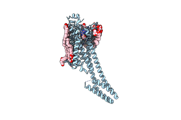 |
In Meso Structure Of The Adenosine A2A G Protein-Coupled Receptor, A2Ar, In 7.10 Monoacylglycerol
Organism: Homo sapiens
Method: X-RAY DIFFRACTION Resolution:2.37 Å Release Date: 2024-04-03 Classification: MEMBRANE PROTEIN Ligands: ZMA, CLR, A1H2K, A1H52, GOL, NA |
 |
In Meso Structure Of Apolipoprotein N-Acyltransferase, Lnt, From Escherichia Coli In 7.10 Monoacylglycerol
Organism: Escherichia coli
Method: X-RAY DIFFRACTION Resolution:2.19 Å Release Date: 2024-04-03 Classification: MEMBRANE PROTEIN Ligands: A1H2K, GOL |
 |
Organism: Rattus norvegicus
Method: ELECTRON MICROSCOPY Release Date: 2024-02-07 Classification: MEMBRANE PROTEIN Ligands: PEX |
 |
Organism: Rattus norvegicus
Method: ELECTRON MICROSCOPY Release Date: 2024-02-07 Classification: MEMBRANE PROTEIN Ligands: PEX, FZ4, R2R |
 |
Organism: Oryctolagus cuniculus
Method: ELECTRON MICROSCOPY Release Date: 2024-02-07 Classification: MEMBRANE PROTEIN Ligands: R2R, POV, ERG |
 |
Wildtype Rabbit Trpv5 Into Nanodiscs In The Presence Of Pi(4,5)P2 And Ruthenium Red
Organism: Oryctolagus cuniculus
Method: ELECTRON MICROSCOPY Release Date: 2024-02-07 Classification: MEMBRANE PROTEIN Ligands: ERG, R2R, CPL |