
Search Count: 140
 |
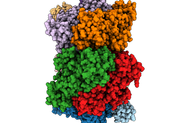
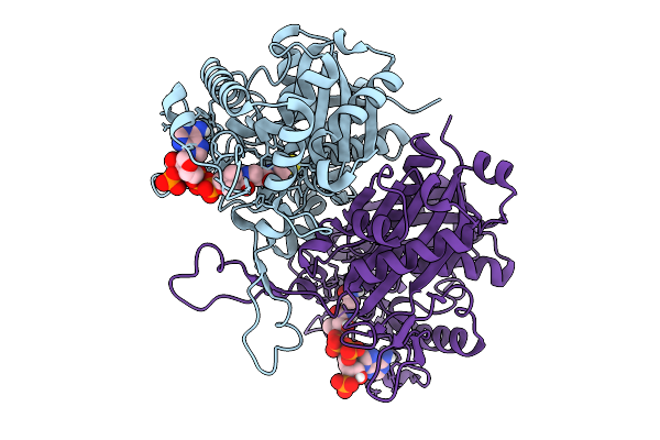
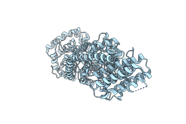
Organism: Methanococcoides burtonii
Method: X-RAY DIFFRACTION Release Date: 2025-11-19 Classification: LIGASE Ligands: MG |
 |
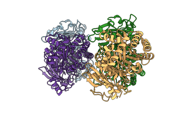
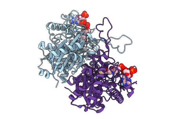
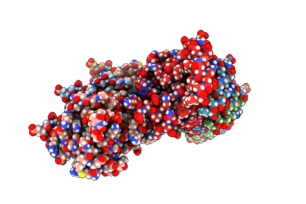
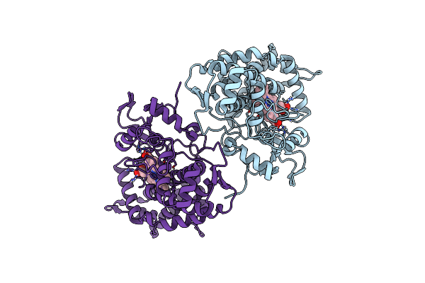
Crystal Structure Of Rubisco In Complex With Cabp From Methanococcoides Burtonii
Organism: Methanococcoides burtonii
Method: X-RAY DIFFRACTION Release Date: 2025-11-19 Classification: LIGASE Ligands: CAP, MG |
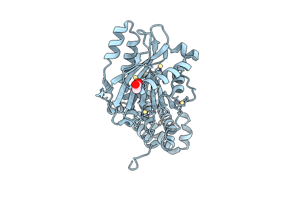 |
Organism: Acinetobacter baylyi adp1
Method: X-RAY DIFFRACTION Release Date: 2025-11-12 Classification: PROTEIN BINDING Ligands: ACT, CD |
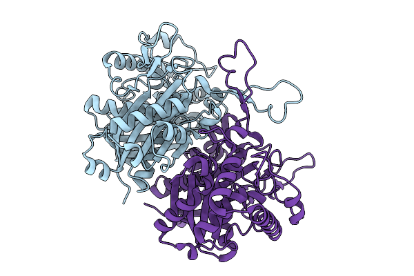 |
Organism: Burkholderia sp. rf2-non_bp3
Method: X-RAY DIFFRACTION Release Date: 2025-08-27 Classification: LYASE |
 |
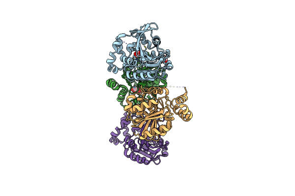
Crystal Structure Of Polyketoacyl-Coa Thiolase From Burkholderia Sp In Complex With Butyryl-Coa
Organism: Burkholderia sp. rf2-non_bp3
Method: X-RAY DIFFRACTION Release Date: 2025-08-27 Classification: LYASE Ligands: COA |
 |
Crystal Structure Of Polyketoacyl-Coa Thiolase From Burkholderia Sp. In Complex With Acetyl-Coa
Organism: Burkholderia sp. rf2-non_bp3
Method: X-RAY DIFFRACTION Release Date: 2025-08-27 Classification: LYASE Ligands: COA |
 |
Crystal Structure Of Polyketoacyl-Coa Thiolase From Burkholderia Sp. In Complex With Acetoacetyl-Coa
Organism: Burkholderia sp. rf2-non_bp3
Method: X-RAY DIFFRACTION Release Date: 2025-08-27 Classification: LYASE Ligands: COA, A1BNQ |
 |
Organism: Enterobacteria phage prd1
Method: X-RAY DIFFRACTION Resolution:1.70 Å Release Date: 2025-04-09 Classification: DNA BINDING PROTEIN |
 |
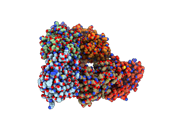
Enterobacteriaphage Prd1 - P12 Protein Filament In Complex With Poly(Dt) Ssdna
Organism: Enterobacteria phage prd1, Synthetic construct
Method: ELECTRON MICROSCOPY Resolution:2.75 Å Release Date: 2025-03-19 Classification: REPLICATION |
 |
Crystal Structure Of Polyketide Synthase (Pks) Thioreductase Domain From Streptomyces Coelicolor
Organism: Streptomyces coelicolor
Method: X-RAY DIFFRACTION Release Date: 2024-11-27 Classification: BIOSYNTHETIC PROTEIN Ligands: NDP, GOL, BTB |
 |
Organism: Grindelia hirsutula
Method: X-RAY DIFFRACTION Resolution:2.12 Å Release Date: 2024-11-06 Classification: PLANT PROTEIN |
 |
Organism: Uncultured proteobacterium
Method: X-RAY DIFFRACTION Resolution:1.90 Å Release Date: 2024-10-23 Classification: HYDROLASE Ligands: GOL |
 |
Crystal Structure Of Chms Dehydrogenase Pmdc From Comamonas Testosteroni Bound To Cofactor Nadp
Organism: Comamonas testosteroni atcc 11996
Method: X-RAY DIFFRACTION Resolution:2.34 Å Release Date: 2024-09-18 Classification: OXIDOREDUCTASE Ligands: SO4, NAP |
 |
Organism: Pilosella officinarum
Method: X-RAY DIFFRACTION Resolution:2.36 Å Release Date: 2024-08-07 Classification: TRANSFERASE |
 |
Organism: Synthetic construct
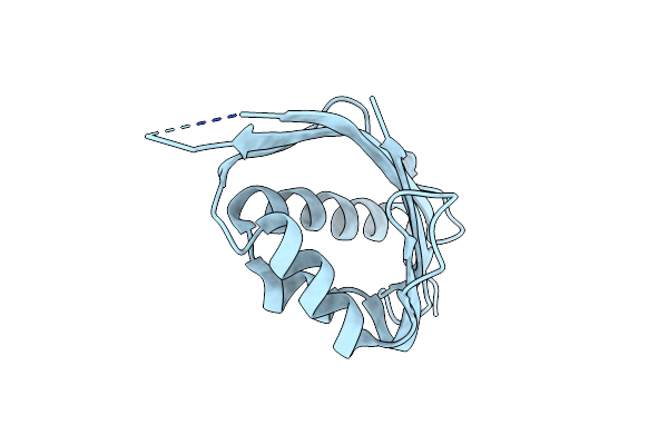
Method: X-RAY DIFFRACTION Release Date: 2024-05-22 Classification: DE NOVO PROTEIN |
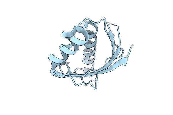 |
Organism: Synthetic construct
Method: X-RAY DIFFRACTION Release Date: 2024-05-22 Classification: DE NOVO PROTEIN |
 |
Organism: Metagenome
Method: X-RAY DIFFRACTION Resolution:1.53 Å Release Date: 2023-05-10 Classification: BIOSYNTHETIC PROTEIN Ligands: HEM |
 |
Organism: Populus trichocarpa
Method: X-RAY DIFFRACTION Resolution:2.37 Å Release Date: 2023-03-15 Classification: TRANSFERASE Ligands: NAG |
 |
Crystal Structure Of Gals1 In Complex With Manganese From Populus Trichocarpas
Organism: Populus trichocarpa
Method: X-RAY DIFFRACTION Resolution:2.56 Å Release Date: 2023-03-15 Classification: TRANSFERASE Ligands: MN, NAG |
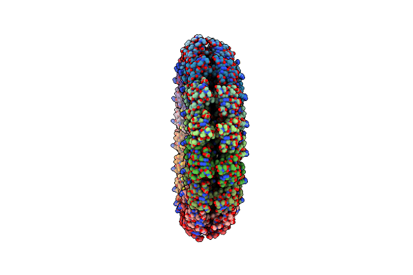 |
An Asymmetric Disk Assembly Formed By Tandem Dimers Of The Tobacco Mosaic Viral Capsid Protein (Tmv)
Organism: Tobacco mosaic virus (vulgare)
Method: X-RAY DIFFRACTION Resolution:2.80 Å Release Date: 2023-03-01 Classification: VIRAL PROTEIN |