
Search Count: 16
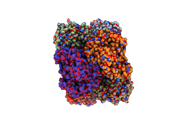 |
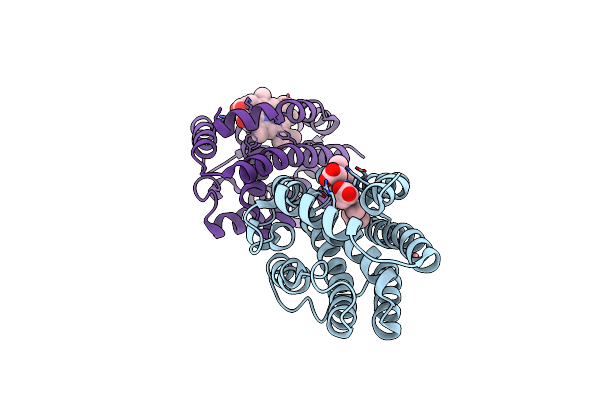
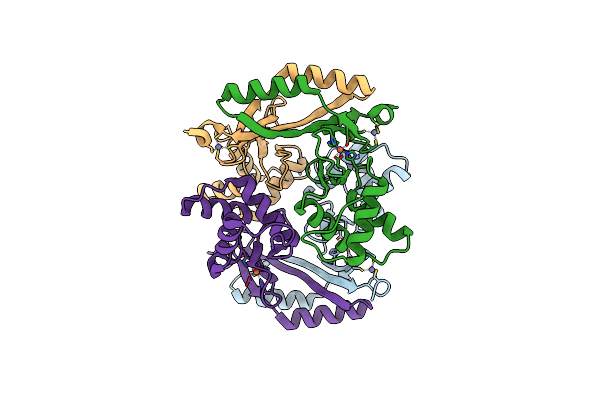
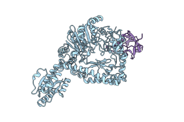
Crystal Structure Of The Recombinant Codh From Rhodopspirillum Rubrum Produced In Escherichia Coli
Organism: Rhodospirillum rubrum
Method: X-RAY DIFFRACTION Resolution:2.90 Å Release Date: 2025-02-19 Classification: OXIDOREDUCTASE Ligands: SF4, RQM, NA |
 |
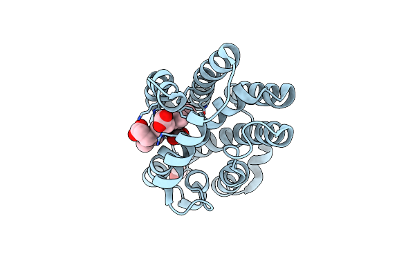
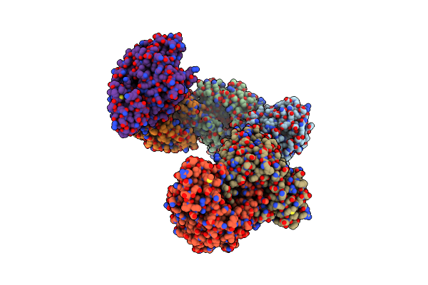
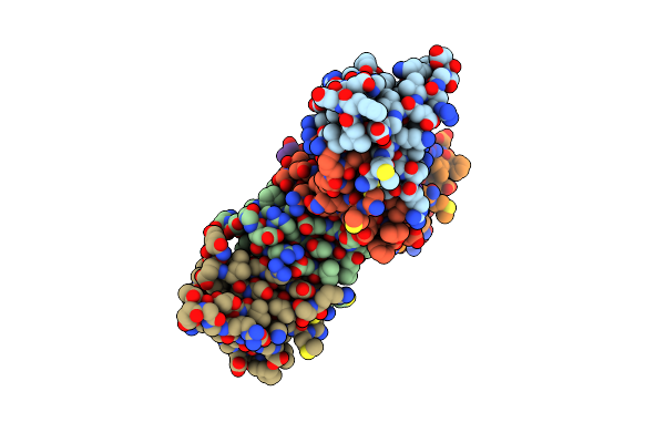
Crystal Structure Of Heme-Oxygenase From Corynebacterium Diphtheriae Complexed With Cobalt-Porphyrine (Humo-Co(Iii))
Organism: Corynebacterium diphtheriae
Method: X-RAY DIFFRACTION Resolution:1.15 Å Release Date: 2024-10-16 Classification: OXIDOREDUCTASE Ligands: COH, GOL, EDO, CIT, CL |
 |
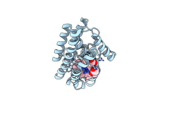
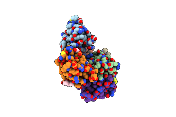
Crystal Structure Of Heme-Oxygenase From Corynebacterium Diphtheriae Complexed With Cobalt-Porphyrine (Humo-Co(Iii)) Flash-Cooled Under Co2 Pressure
Organism: Corynebacterium diphtheriae
Method: X-RAY DIFFRACTION Resolution:1.85 Å Release Date: 2024-10-16 Classification: OXIDOREDUCTASE Ligands: COH, EDO, CL, CO2 |
 |
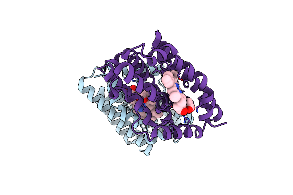
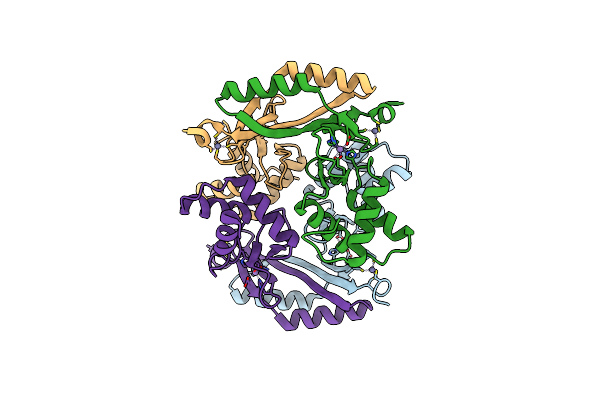
Crystal Structure Of Heme-Oxygenase Mutant G139A From Corynebacterium Diphtheriae Complexed With Cobalt-Porphyrine (Humo-Co(Iii))
Organism: Corynebacterium diphtheriae
Method: X-RAY DIFFRACTION Resolution:1.40 Å Release Date: 2024-10-16 Classification: OXIDOREDUCTASE Ligands: COH, CL |
 |
Crystal Structure Of Heme-Oxygenase Mutant H20C From Corynebacterium Diphtheriae Complexed With Cobalt-Porphyrine (Humo-Co(Iii))
Organism: Corynebacterium diphtheriae
Method: X-RAY DIFFRACTION Resolution:2.30 Å Release Date: 2024-10-16 Classification: OXIDOREDUCTASE Ligands: COH |
 |
Crystal Structure Of Heme-Oxygenase Mutant I143K From Corynebacterium Diphtheriae Complexed With Cobalt-Porphyrine (Humo-Co(Iii))
Organism: Corynebacterium diphtheriae
Method: X-RAY DIFFRACTION Resolution:2.84 Å Release Date: 2024-10-16 Classification: OXIDOREDUCTASE Ligands: CO, COH, CL |
 |
Crystal Structure Of The Hexxh Domain Of Chlbhexxh A Novel Alpha-Ketoglutarate Dependent Oxygenase
Organism: Chlorogloeopsis sp.
Method: X-RAY DIFFRACTION Resolution:2.80 Å Release Date: 2024-10-02 Classification: OXIDOREDUCTASE Ligands: PO4 |
 |
Structure Of Ferric Uptake Regulator From Pseudomonas Aeruginosa With Manganese.
Organism: pseudomonas aeruginosa pao1
Method: X-RAY DIFFRACTION Resolution:2.34 Å Release Date: 2019-07-31 Classification: TRANSCRIPTION Ligands: MN, ZN |
 |
X-Ray Structure Of A Truncated Mutant Of The Metallochaperone Cooj With A High-Affinity Nickel-Binding Site
Organism: Rhodospirillum rubrum
Method: X-RAY DIFFRACTION Resolution:2.04 Å Release Date: 2019-03-27 Classification: METAL BINDING PROTEIN Ligands: NI, PEG, CL, CA, PGE, Z3P |
 |
Organism: Carboxydothermus hydrogenoformans (strain atcc baa-161 / dsm 6008 / z-2901)
Method: X-RAY DIFFRACTION Resolution:2.00 Å Release Date: 2018-11-28 Classification: METAL BINDING PROTEIN Ligands: MPD, GOL |
 |
Organism: Francisella tularensis
Method: X-RAY DIFFRACTION Resolution:1.70 Å Release Date: 2018-05-16 Classification: DNA BINDING PROTEIN Ligands: MN, ZN |
 |
Organism: Francisella tularensis
Method: X-RAY DIFFRACTION Resolution:1.80 Å Release Date: 2018-05-16 Classification: TRANSCRIPTION Ligands: FE, ZN |
 |
Crystal Structure Of The Apo-Form Of The Co Dehydrogenase Accessory Protein Coot From Rhodospirillum Rubrum
Organism: Rhodospirillum rubrum
Method: X-RAY DIFFRACTION Resolution:1.90 Å Release Date: 2017-05-10 Classification: nickel-binding protein |
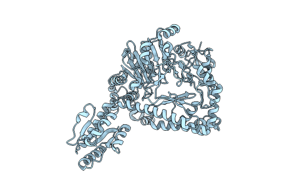 |
Organism: Escherichia coli k-12
Method: ELECTRON MICROSCOPY Resolution:6.10 Å Release Date: 2016-09-21 Classification: LYASE |
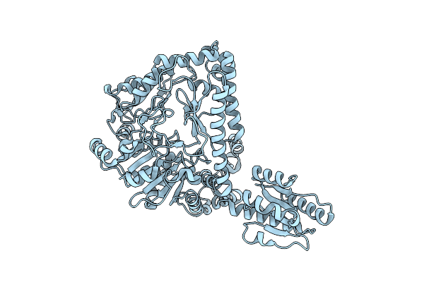 |
Organism: Escherichia coli
Method: ELECTRON MICROSCOPY Resolution:5.50 Å Release Date: 2016-09-21 Classification: LYASE |
 |
Revisited Cryo-Em Structure Of Inducible Lysine Decarboxylase Complexed With Lara Domain Of Rava Atpase
Organism: Escherichia coli k-12
Method: ELECTRON MICROSCOPY Resolution:6.20 Å Release Date: 2016-09-21 Classification: HYDROLASE/ISOMERASE |