
Search Count: 63
All
Selected
 |
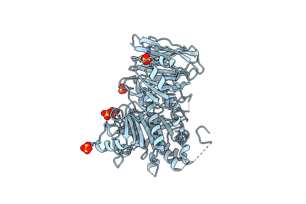
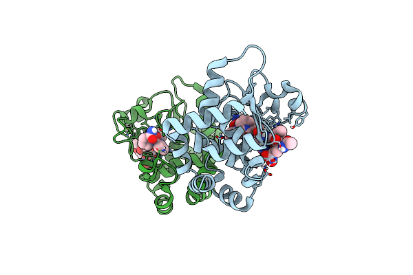
Crystal Structure Of The Accessory Translocation Atpase, Seca2, From Clostridium Difficile, In Complex With Adenosine-5'-(Gamma-Thio)-Triphosphate
Organism: Peptoclostridium difficile (strain 630)
Method: X-RAY DIFFRACTION Resolution:2.90 Å Release Date: 2020-11-18 Classification: PROTEIN TRANSPORT Ligands: AGS |
 |
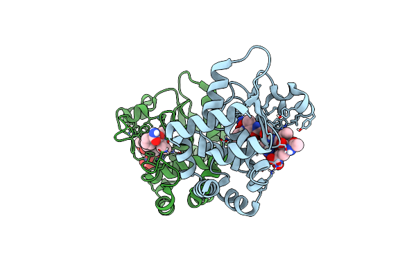
Crystal Structure Of The Accessory Translocation Atpase, Seca2, From Clostridium Difficile
Organism: Peptoclostridium difficile (strain 630)
Method: X-RAY DIFFRACTION Resolution:2.30 Å Release Date: 2020-10-07 Classification: PROTEIN TRANSPORT |
 |
Crystal Structure Of The Chitinase Domain Of The Spore Coat Protein Cote From Clostridium Difficile
Organism: Peptoclostridium difficile (strain 630), Escherichia coli k-12
Method: X-RAY DIFFRACTION Resolution:1.30 Å Release Date: 2020-07-22 Classification: STRUCTURAL PROTEIN Ligands: 1PE, PEG |
 |
Organism: Peptoclostridium difficile 630
Method: X-RAY DIFFRACTION Resolution:2.60 Å Release Date: 2020-02-19 Classification: HYDROLASE |
 |
1.85 Angstrom Resolution Crystal Structure Of Class D Beta-Lactamase From Clostridium Difficile 630
Organism: Peptoclostridium difficile (strain 630)
Method: X-RAY DIFFRACTION Resolution:1.85 Å Release Date: 2019-12-25 Classification: HYDROLASE Ligands: PGE, PEG, PPI, GOL |
 |
Crystal Structure Of Mura From Clostridium Difficile In The Presence Of Udp-N-Acetyl-Alpha-D-Muramic Acid With Modified Cys116 (S-[(1S)-1-Carboxy-1-(Phosphonooxy)Ethyl]-L-Cysteine)
Organism: Peptoclostridium difficile (strain 630)
Method: X-RAY DIFFRACTION Resolution:1.70 Å Release Date: 2019-11-27 Classification: TRANSFERASE Ligands: EPZ, EDO |
 |
Crystal Structure Of Mura From Clostridium Difficile, Mutation C116D, N The Presence Of Udp-N-Acetylmuramic Acid
Organism: Peptoclostridium difficile (strain 630)
Method: X-RAY DIFFRACTION Resolution:1.65 Å Release Date: 2019-11-27 Classification: TRANSFERASE Ligands: EDO, EPZ |
 |
Crystal Structure Of Mura From Clostridium Difficile, Mutant C116S, In The Presence Of Uridine-Diphosphate-N-Acetylglucosamine
Organism: Peptoclostridium difficile (strain 630)
Method: X-RAY DIFFRACTION Resolution:1.70 Å Release Date: 2019-11-27 Classification: TRANSFERASE Ligands: EDO, UD1 |
 |
Crystal Structure Of Mura From Clostridium Difficile, Mutation C116S, In The Presence Of Uridine-Diphosphate-2(N-Acetylglucosaminyl) Butyric Acid
Organism: Peptoclostridium difficile (strain 630)
Method: X-RAY DIFFRACTION Resolution:1.80 Å Release Date: 2019-11-27 Classification: TRANSFERASE Ligands: EPU, EDO, NA |
 |
The Ligand-Bound, Closed Structure Of Cd0873, A Substrate Binding Protein With Adhesive Properties From Clostridium Difficile.
Organism: Peptoclostridium difficile 630
Method: X-RAY DIFFRACTION Resolution:1.35 Å Release Date: 2019-08-28 Classification: TRANSPORT PROTEIN Ligands: TYR, EDO, PEG |
 |
The Ligand-Bound, Open Structure Of Cd0873, A Substrate Binding Protein With Adhesive Properties From Clostridium Difficile.
Organism: Peptoclostridium difficile 630
Method: X-RAY DIFFRACTION Resolution:1.80 Å Release Date: 2019-08-28 Classification: TRANSPORT PROTEIN Ligands: EDO, PGE, PEG |
 |
The Ligand-Free, Open Structure Of Cd0873, A Substrate Binding Protein With Adhesive Properties From Clostridium Difficile.
Organism: Peptoclostridium difficile 630
Method: X-RAY DIFFRACTION Resolution:2.50 Å Release Date: 2019-08-28 Classification: TRANSPORT PROTEIN |
 |
Organism: Peptoclostridium difficile (strain r20291)
Method: X-RAY DIFFRACTION Resolution:1.55 Å Release Date: 2019-06-26 Classification: SIGNALING PROTEIN Ligands: SO4 |
 |
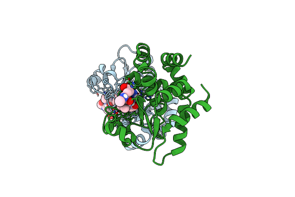
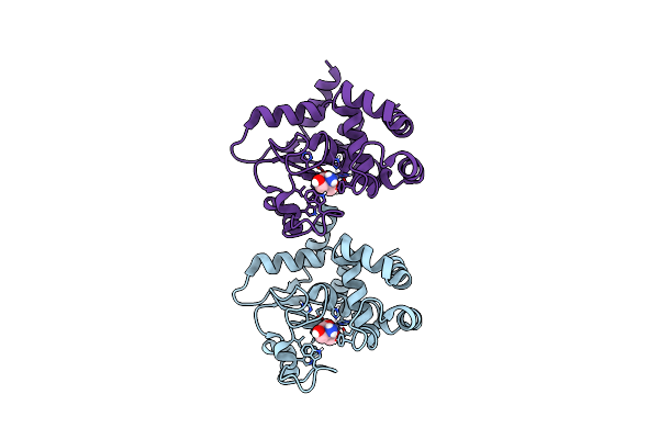
Crystal Structure Of Apo Ppep-1(E143A/Y178F) In Complex With Substrate Peptide Ac-Evnapvp-Conh2
Organism: Peptoclostridium difficile, Clostridioides difficile
Method: X-RAY DIFFRACTION Resolution:1.39 Å Release Date: 2019-06-12 Classification: HYDROLASE Ligands: ZN |
 |
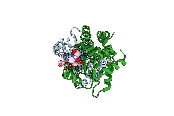
Crystal Structure Of Ppep-1(E143A/Y178F) In Complex With Substrate Peptide Ac-Evnpavp-Conh2
Organism: Peptoclostridium difficile, Clostridioides difficile
Method: X-RAY DIFFRACTION Resolution:1.83 Å Release Date: 2019-06-12 Classification: HYDROLASE Ligands: ZN |
 |
Crystal Structure Of Holo Ppep-1(E143A/Y178F) In Complex With Product Peptide Ac-Evnp-Co2 (Substrate Peptide: Ac-Evnpavp-Conh2)
Organism: Peptoclostridium difficile, Clostridioides difficile
Method: X-RAY DIFFRACTION Resolution:1.45 Å Release Date: 2019-06-12 Classification: HYDROLASE Ligands: ZN |
 |
Crystal Structure Of Holo Ppep-1(E143A/Y178F) In Complex With Product Peptide Ac-Evnp-Co2 (Substrate Peptide: Ac-Evnppvp-Conh2)
Organism: Peptoclostridium difficile, Clostridioides difficile
Method: X-RAY DIFFRACTION Resolution:1.05 Å Release Date: 2019-06-12 Classification: HYDROLASE Ligands: ZN, NI |
 |
Crystal Structure Of Holo Ppep-1(E143A/Y178F) In Complex With Substrate Peptide Ac-Evnapvp-Conh2
Organism: Peptoclostridium difficile , Clostridioides difficile
Method: X-RAY DIFFRACTION Resolution:1.81 Å Release Date: 2019-06-12 Classification: HYDROLASE Ligands: ZN |
 |
Crystal Structure Of Apo Ppep-1(E143A/Y178F) In Complex With Fibrinogen-Derived Substrate Peptide Ac-Slrpapp-Conh2
Organism: Peptoclostridium difficile, Homo sapiens
Method: X-RAY DIFFRACTION Resolution:1.94 Å Release Date: 2019-06-12 Classification: HYDROLASE Ligands: PO4 |
 |
Organism: Peptoclostridium difficile
Method: X-RAY DIFFRACTION Resolution:2.02 Å Release Date: 2019-06-12 Classification: HYDROLASE Ligands: TRS, ZN |