
Search Count: 177
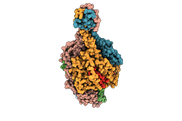 |
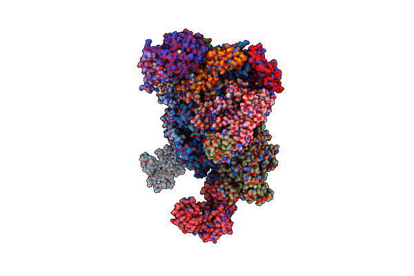
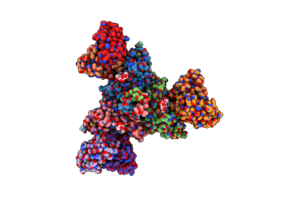
Respiratory Syncytial Virus Pre-F Trimer Bound By Neutralizing Antibody Pr306007
Organism: Respiratory syncytial virus a2, Homo sapiens
Method: ELECTRON MICROSCOPY Release Date: 2025-11-12 Classification: STRUCTURAL PROTEIN/IMMUNE SYSTEM |
 |
Organism: Escherichia coli
Method: X-RAY DIFFRACTION Release Date: 2025-09-17 Classification: FLUORESCENT PROTEIN Ligands: NO3, SO4 |
 |
Organism: Escherichia coli
Method: X-RAY DIFFRACTION Release Date: 2025-09-17 Classification: FLUORESCENT PROTEIN |
 |
Organism: Homo sapiens
Method: X-RAY DIFFRACTION Release Date: 2025-09-10 Classification: APOPTOSIS Ligands: MG, PD |
 |
 |
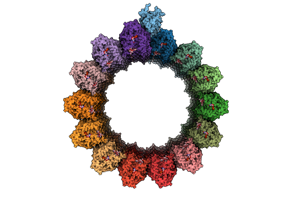
Organism: Tetrahymena thermophila cu428
Method: ELECTRON MICROSCOPY Release Date: 2025-07-30 Classification: PROTEIN BINDING Ligands: GTP, MG, GDP |
 |
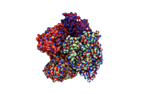
Organism: Bos taurus
Method: ELECTRON MICROSCOPY Release Date: 2025-07-30 Classification: STRUCTURAL PROTEIN Ligands: GTP, MG, GDP, TA1 |
 |
Organism: Pseudomonas aeruginosa
Method: X-RAY DIFFRACTION Release Date: 2025-07-23 Classification: DNA BINDING PROTEIN |
 |
Organism: Homo sapiens, Recombinant vesicular stomatitis indiana virus rvsv-g/gfp, Bos taurus
Method: ELECTRON MICROSCOPY Release Date: 2025-07-23 Classification: PROTEIN BINDING Ligands: GTP, MG, GDP, TA1 |
 |
Organism: Bacillus subtilis (strain 168)
Method: X-RAY DIFFRACTION Release Date: 2025-04-30 Classification: OXIDOREDUCTASE Ligands: HEM, PLM, GOL, SO4, PEG |
 |
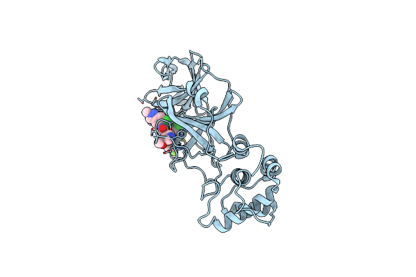
Organism: Severe acute respiratory syndrome coronavirus 2
Method: X-RAY DIFFRACTION Resolution:1.92 Å Release Date: 2025-04-02 Classification: VIRAL PROTEIN Ligands: A1EGN |
 |
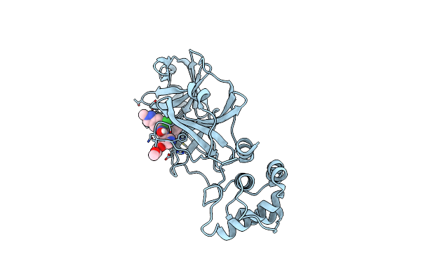
Organism: Severe acute respiratory syndrome coronavirus 2
Method: X-RAY DIFFRACTION Resolution:1.91 Å Release Date: 2025-04-02 Classification: VIRAL PROTEIN Ligands: A1EGQ |
 |
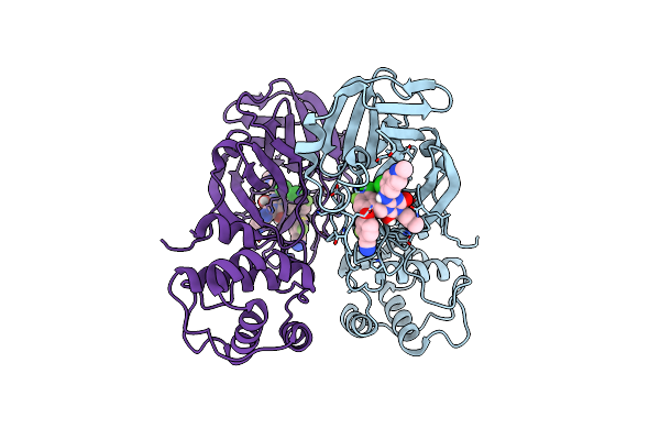
Organism: Severe acute respiratory syndrome coronavirus 2
Method: X-RAY DIFFRACTION Resolution:2.30 Å Release Date: 2025-04-02 Classification: VIRAL PROTEIN Ligands: A1EGP |
 |
Organism: Severe acute respiratory syndrome coronavirus 2
Method: X-RAY DIFFRACTION Resolution:1.67 Å Release Date: 2025-04-02 Classification: VIRAL PROTEIN Ligands: A1EGR |
 |
Organism: Severe acute respiratory syndrome coronavirus 2
Method: X-RAY DIFFRACTION Resolution:2.45 Å Release Date: 2025-04-02 Classification: VIRAL PROTEIN Ligands: A1EGS |
 |
Organism: Pipistrellus bat coronavirus hku5, Homo sapiens
Method: ELECTRON MICROSCOPY Release Date: 2025-03-12 Classification: VIRAL PROTEIN/HYDROLASE Ligands: NAG |
 |
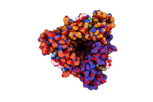
Respiratory Syncytial Virus Fusion Protein In The Postfusion Conformation In Complex With Monoclonal Antibody 131-2A Fab
Organism: Human respiratory syncytial virus, Mus musculus
Method: ELECTRON MICROSCOPY Release Date: 2025-02-26 Classification: VIRAL PROTEIN |
 |
Organism: Spinacia oleracea
Method: ELECTRON MICROSCOPY Release Date: 2024-12-04 Classification: TRANSCRIPTION Ligands: FE |
 |
Organism: Measles morbillivirus, Mus musculus
Method: ELECTRON MICROSCOPY Release Date: 2024-07-03 Classification: VIRAL PROTEIN Ligands: NAG |
 |
Structure Of The Measles Virus Fusion Protein In The Post-Fusion Conformation
Organism: Measles virus strain ichinose-b95a
Method: ELECTRON MICROSCOPY Release Date: 2024-07-03 Classification: VIRAL PROTEIN Ligands: NAG |