Search Count: 38
 |
Structural Architecture Of The Acidic Region Of The B Domain Of Coagulation Factor V
Organism: Homo sapiens
Method: ELECTRON MICROSCOPY Release Date: 2023-10-04 Classification: BLOOD CLOTTING Ligands: NAG |
 |
Organism: Homo sapiens
Method: ELECTRON MICROSCOPY Release Date: 2023-03-15 Classification: BLOOD CLOTTING |
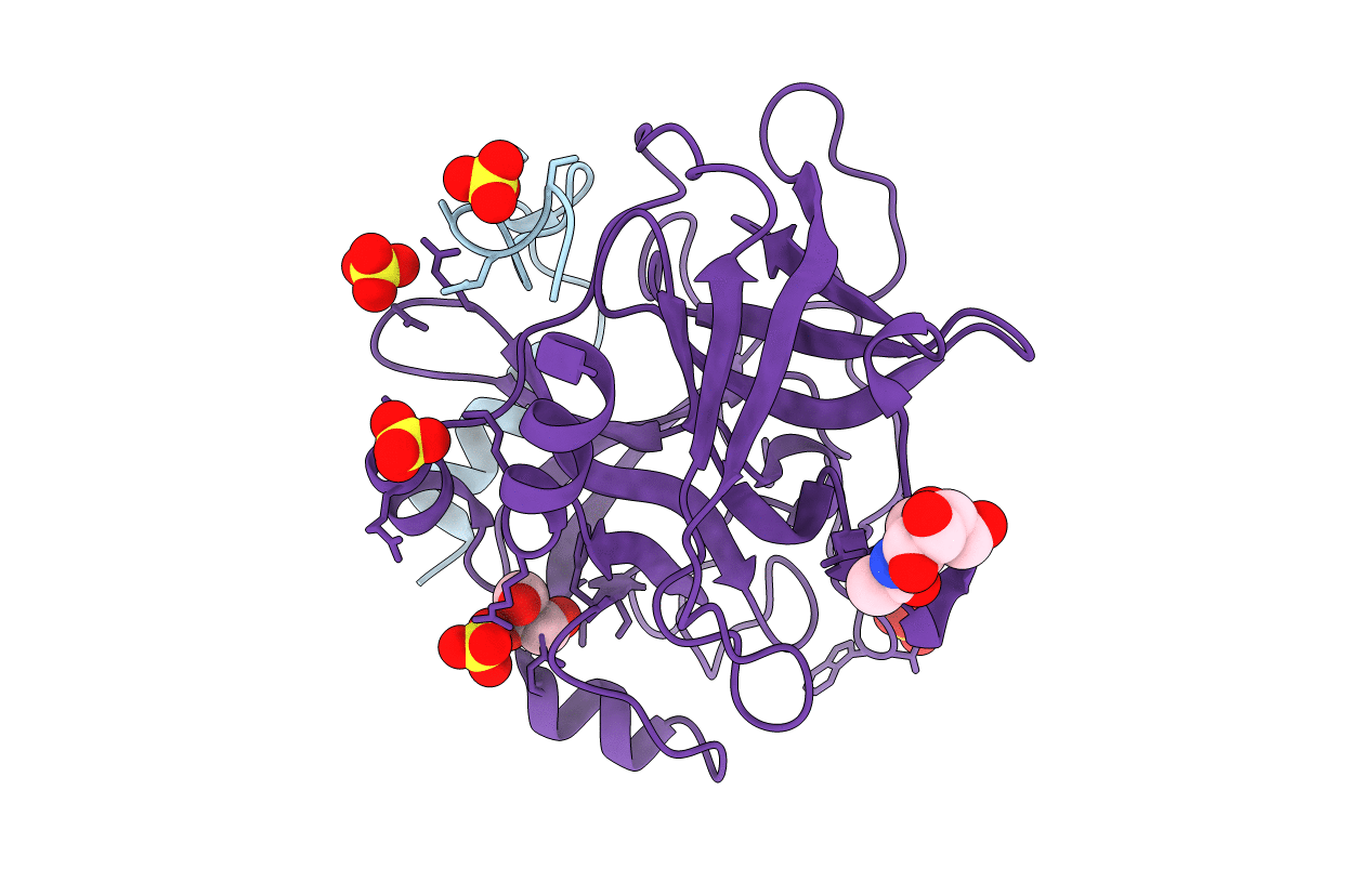 |
Human Alpha-Thrombin With 180- And 220- Loops Replaced With Homologous Loops From Protein C
Organism: Homo sapiens
Method: X-RAY DIFFRACTION Resolution:2.10 Å Release Date: 2021-12-08 Classification: BLOOD CLOTTING Ligands: SO4, NAG, GOL |
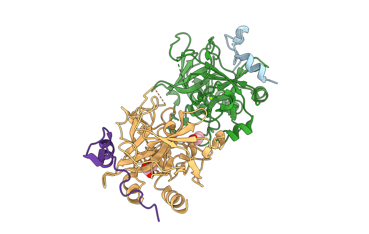 |
Organism: Homo sapiens
Method: X-RAY DIFFRACTION Resolution:1.70 Å Release Date: 2019-12-18 Classification: HYDROLASE Ligands: GOL, MG |
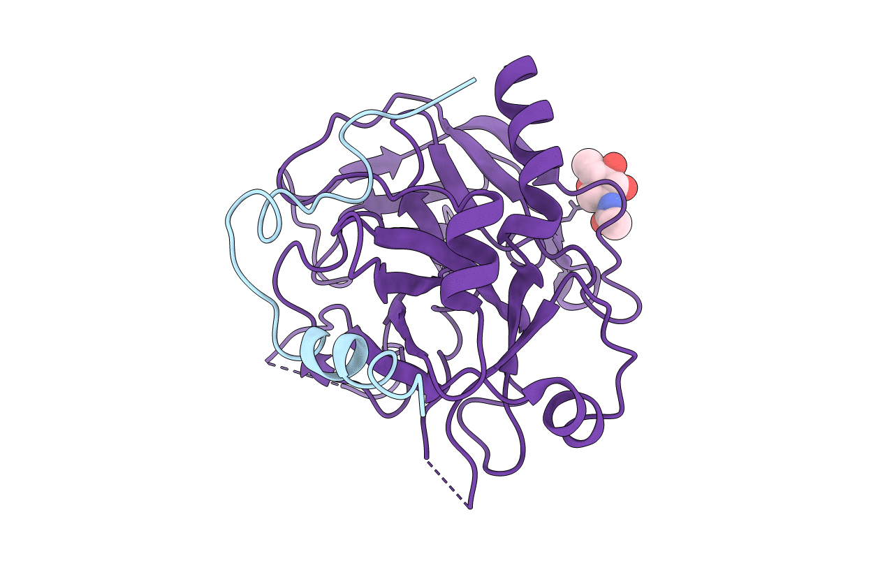 |
Organism: Homo sapiens
Method: X-RAY DIFFRACTION Resolution:2.80 Å Release Date: 2019-12-18 Classification: HYDROLASE Ligands: NAG |
 |
Organism: Homo sapiens
Method: X-RAY DIFFRACTION Resolution:3.30 Å Release Date: 2019-09-04 Classification: HYDROLASE Ligands: ZN, NAG |
 |
Organism: Homo sapiens
Method: X-RAY DIFFRACTION Resolution:2.40 Å Release Date: 2019-09-04 Classification: hydrolase/hydrolase inhibitor Ligands: ZN, 0G6, NA |
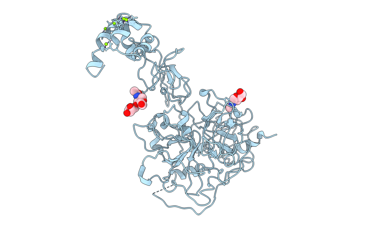 |
Organism: Homo sapiens
Method: X-RAY DIFFRACTION Resolution:6.00 Å Release Date: 2018-06-27 Classification: HYDROLASE Ligands: MG, NAG |
 |
Organism: Homo sapiens
Method: X-RAY DIFFRACTION Resolution:4.12 Å Release Date: 2018-02-28 Classification: BLOOD CLOTTING Ligands: MG, NAG |
 |
Crystal Structure Of Thrombin Mutant W215A/E217A Fused To Egf456 Of Thrombomodulin Via A 31-Residue Linker And Bound To Ppack
Organism: Homo sapiens
Method: X-RAY DIFFRACTION Resolution:2.34 Å Release Date: 2017-03-29 Classification: HYDROLASE Ligands: 0G6, K, NA, NAG |
 |
Organism: Homo sapiens
Method: X-RAY DIFFRACTION Resolution:1.70 Å Release Date: 2015-03-11 Classification: HYDROLASE Ligands: K, GOL |
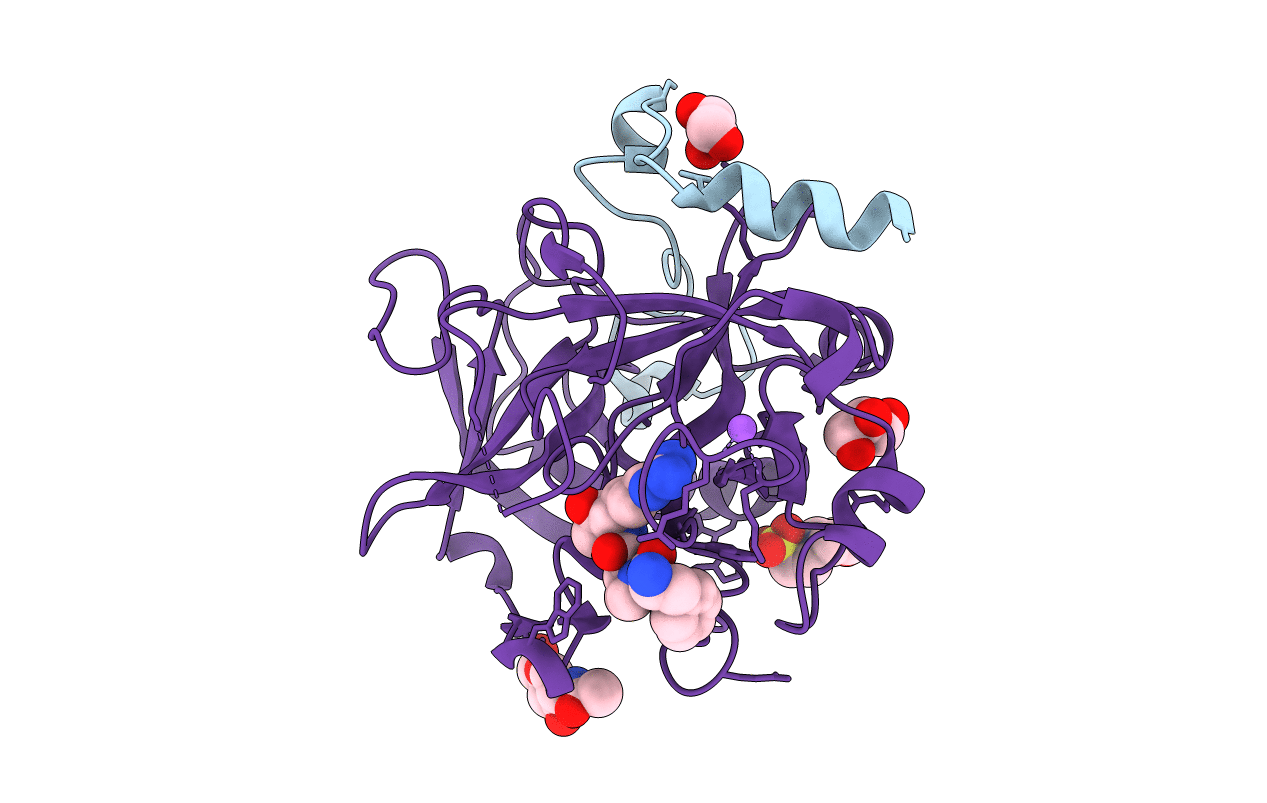 |
Organism: Homo sapiens
Method: X-RAY DIFFRACTION Resolution:1.84 Å Release Date: 2015-03-11 Classification: HYDROLASE/HYDROLASE INHIBITOR Ligands: 0G6, MES, GOL, NA, NAG |
 |
Structure Of Prethrombin-2 Mutant S195A Bound To The Active Site Inhibitor Argatroban
Organism: Homo sapiens
Method: X-RAY DIFFRACTION Resolution:3.00 Å Release Date: 2014-11-05 Classification: HYDROLASE/HYDROLASE INHIBITOR Ligands: 15U |
 |
Organism: Homo sapiens
Method: X-RAY DIFFRACTION Resolution:2.81 Å Release Date: 2014-05-21 Classification: HYDROLASE Ligands: NAG |
 |
Organism: Homo sapiens
Method: X-RAY DIFFRACTION Resolution:3.38 Å Release Date: 2014-05-21 Classification: HYDROLASE Ligands: CA, NAG |
 |
Organism: Homo sapiens
Method: X-RAY DIFFRACTION Resolution:2.19 Å Release Date: 2013-03-13 Classification: HYDROLASE/PEPTIDE |
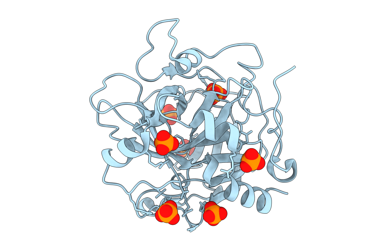 |
Organism: Homo sapiens
Method: X-RAY DIFFRACTION Resolution:2.40 Å Release Date: 2013-03-13 Classification: HYDROLASE Ligands: PO4 |
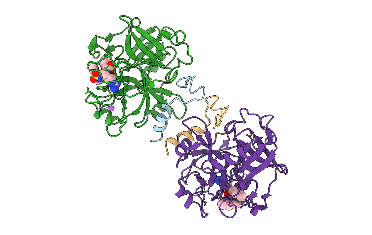 |
Structure Of Thrombin Mutant S195A Bound To The Active Site Inhibitor Argatroban
Organism: Homo sapiens
Method: X-RAY DIFFRACTION Resolution:2.40 Å Release Date: 2013-03-13 Classification: HYDROLASE/HYDROLASE INHIBITOR |
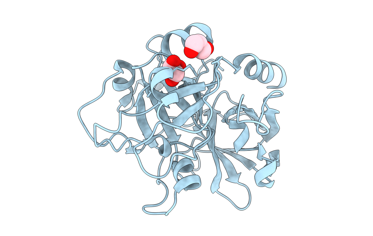 |
Organism: Homo sapiens
Method: X-RAY DIFFRACTION Resolution:1.90 Å Release Date: 2011-11-30 Classification: HYDROLASE Ligands: GOL |
 |
Organism: Homo sapiens
Method: X-RAY DIFFRACTION Resolution:2.20 Å Release Date: 2011-11-30 Classification: HYDROLASE |

