
Search Count: 172
 |
Organism: Oscillatoria acuminata
Method: X-RAY DIFFRACTION Resolution:1.95 Å Release Date: 2025-02-26 Classification: LYASE Ligands: FMN, GOL, CA |
 |
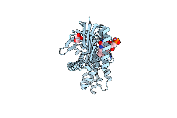
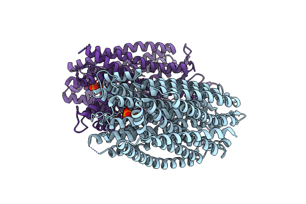
Organism: Homo sapiens
Method: ELECTRON MICROSCOPY Release Date: 2025-02-05 Classification: DE NOVO PROTEIN Ligands: NAG |
 |
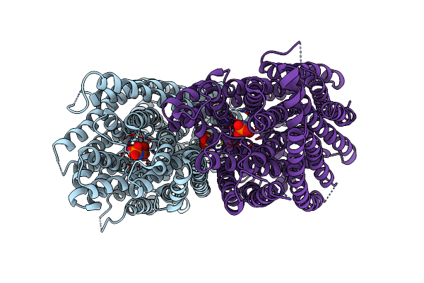
Organism: Homo sapiens
Method: ELECTRON MICROSCOPY Release Date: 2025-02-05 Classification: DE NOVO PROTEIN Ligands: NAG |
 |
Organism: Lobophyllia hemprichii
Method: X-RAY DIFFRACTION Resolution:1.75 Å Release Date: 2024-11-27 Classification: FLUORESCENT PROTEIN Ligands: SO4 |
 |
Structure Of Rskiiro Using Ssx After Illumination With 0.1 Mj/Mm^2 Of 405 Nm Light
Organism: Lobophyllia hemprichii
Method: X-RAY DIFFRACTION Resolution:1.75 Å Release Date: 2024-11-27 Classification: FLUORESCENT PROTEIN Ligands: SO4, GOL |
 |
Structure Of Rskiiro Using Ssx After Illumination With 0.53 Mj/Mm^2 Of 405 Nm Light
Organism: Lobophyllia hemprichii
Method: X-RAY DIFFRACTION Resolution:1.70 Å Release Date: 2024-11-27 Classification: FLUORESCENT PROTEIN Ligands: SO4, GOL |
 |
Structure Of Rskiiro Using Ssx After Illumination With 1.78 Mj/Mm^2 Of 405 Nm Light
Organism: Lobophyllia hemprichii
Method: X-RAY DIFFRACTION Resolution:1.75 Å Release Date: 2024-11-27 Classification: FLUORESCENT PROTEIN Ligands: SO4, GOL |
 |
Structure Of Rskiiro Using Ssx After Illumination With 6.74 Mj/Mm^2 Of 405 Nm Light
Organism: Lobophyllia hemprichii
Method: X-RAY DIFFRACTION Resolution:1.75 Å Release Date: 2024-11-27 Classification: FLUORESCENT PROTEIN Ligands: SO4, GOL |
 |
Structure Of Rskiiro Using Ssx After Illumination With 14.43 Mj/Mm^2 Of 405 Nm Light
Organism: Lobophyllia hemprichii
Method: X-RAY DIFFRACTION Resolution:1.75 Å Release Date: 2024-11-27 Classification: FLUORESCENT PROTEIN Ligands: SO4, GOL |
 |
Organism: Homo sapiens
Method: X-RAY DIFFRACTION Resolution:2.10 Å Release Date: 2024-04-17 Classification: STRUCTURAL PROTEIN |
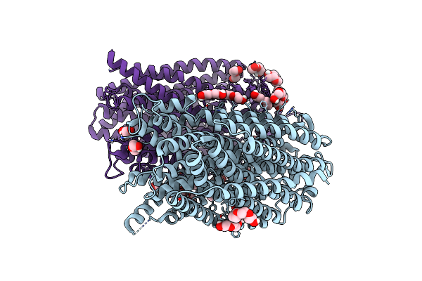 |
Time-Resolved Structure Of K+-Dependent Na+-Ppase From Thermotoga Maritima 0-60-Seconds Post Reaction Initiation With Na+
Organism: Thermotoga maritima
Method: X-RAY DIFFRACTION Resolution:2.59 Å Release Date: 2024-01-17 Classification: MEMBRANE PROTEIN Ligands: LMT, PEG, PGE, PG4 |
 |
Time-Resolved Structure Of K+-Dependent Na+-Ppase From Thermotoga Maritima 300-Seconds Post Reaction Initiation With Na+
Organism: Thermotoga maritima
Method: X-RAY DIFFRACTION Resolution:3.98 Å Release Date: 2024-01-17 Classification: MEMBRANE PROTEIN Ligands: DPO, MG |
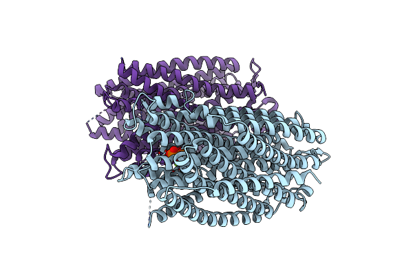 |
Time-Resolved Structure Of K+-Dependent Na+-Ppase From Thermotoga Maritima 600-Seconds Post Reaction Initiation With Na+
Organism: Thermotoga maritima
Method: X-RAY DIFFRACTION Resolution:3.84 Å Release Date: 2024-01-17 Classification: MEMBRANE PROTEIN Ligands: DPO, MG |
 |
Time-Resolved Structure Of K+-Dependent Na+-Ppase From Thermotoga Maritima 3600-Seconds Post Reaction Initiation With Na+
Organism: Thermotoga maritima
Method: X-RAY DIFFRACTION Resolution:4.53 Å Release Date: 2024-01-17 Classification: MEMBRANE PROTEIN Ligands: DPO, MG, K, PO4 |
 |
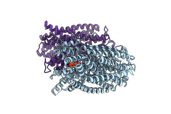
Crystal Structure Of Pyrobaculum Aerophilum Potassium-Independent Proton Pumping Membrane Integral Pyrophosphatase In Complex With Imidodiphosphate And Magnesium, And With Bound Sulphate
Organism: Pyrobaculum aerophilum
Method: X-RAY DIFFRACTION Resolution:3.84 Å Release Date: 2024-01-17 Classification: MEMBRANE PROTEIN Ligands: MG, 2PN, SO4 |
 |
Organism: Homo sapiens
Method: X-RAY DIFFRACTION Resolution:2.00 Å Release Date: 2023-11-29 Classification: STRUCTURAL PROTEIN |
 |
Organism: Homo sapiens
Method: X-RAY DIFFRACTION Resolution:2.00 Å Release Date: 2023-11-01 Classification: STRUCTURAL PROTEIN Ligands: DTT |
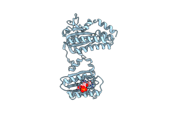 |
Organism: Oscillatoria acuminata pcc 6304
Method: X-RAY DIFFRACTION Resolution:1.50 Å Release Date: 2023-11-01 Classification: LYASE Ligands: FMN, MG |
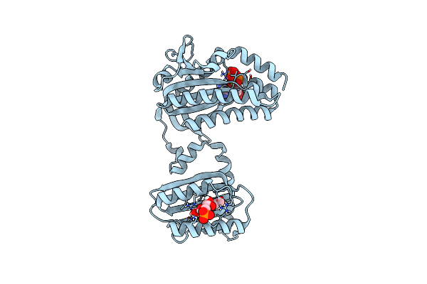 |
Cryogenic Crystal Structure Of The Photoactivated Adenylate Cyclase Oapac With Atp Bound
Organism: Oscillatoria acuminata pcc 6304
Method: X-RAY DIFFRACTION Resolution:2.10 Å Release Date: 2023-11-01 Classification: LYASE Ligands: FMN, ATP, MG |
 |
Cryogenic Crystal Structure Of The Photoactivated Adenylate Cyclase Oapac After 5 Seconds Of Blue Light Illumination
Organism: Oscillatoria acuminata pcc 6304
Method: X-RAY DIFFRACTION Resolution:1.70 Å Release Date: 2023-11-01 Classification: LYASE Ligands: FMN, ATP, MG |