
Search Count: 1,010
 |
Organism: Pseudomonas fluorescens
Method: X-RAY DIFFRACTION Resolution:2.50 Å Release Date: 2023-11-01 Classification: HYDROLASE Ligands: ZIZ, GOL |
 |
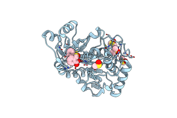
Structure Of The Laspartomycin C Double Mutant G4D D-Allo-Thr9D-Dap In Complex With Geranyl Phosphate
Organism: Streptomyces viridochromogenes
Method: X-RAY DIFFRACTION Resolution:1.04 Å Release Date: 2022-02-02 Classification: ANTIBIOTIC Ligands: CA, RDZ, 9GE |
 |
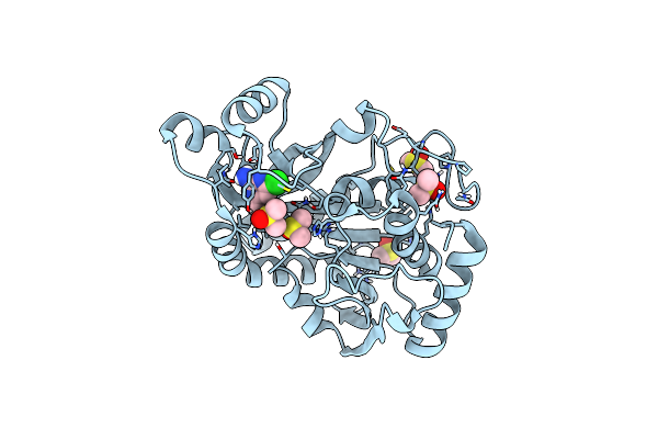
Structure Of The Laspartomycin C Friulimicin-Like Mutant In Complex With Geranyl Phosphate
Organism: Streptomyces viridochromogenes
Method: X-RAY DIFFRACTION Resolution:1.14 Å Release Date: 2022-02-02 Classification: ANTIBIOTIC Ligands: CA, RDZ, CL, CD, NA |
 |
Organism: Homo sapiens, Mus musculus
Method: X-RAY DIFFRACTION Resolution:2.60 Å Release Date: 2021-04-07 Classification: SIGNALING PROTEIN Ligands: EPE, CL |
 |
Organism: Homo sapiens
Method: X-RAY DIFFRACTION Resolution:2.00 Å Release Date: 2020-09-23 Classification: IMMUNE SYSTEM |
 |
Organism: Homo sapiens, Lama glama
Method: X-RAY DIFFRACTION Resolution:1.33 Å Release Date: 2020-09-23 Classification: CELL ADHESION Ligands: PGE, EDO, ACY |
 |
Organism: Lama glama
Method: X-RAY DIFFRACTION Resolution:1.70 Å Release Date: 2020-09-23 Classification: IMMUNE SYSTEM |
 |
Organism: Homo sapiens, Lama glama
Method: X-RAY DIFFRACTION Resolution:2.68 Å Release Date: 2020-09-23 Classification: CELL ADHESION Ligands: GOL, CL |
 |
Organism: Homo sapiens
Method: X-RAY DIFFRACTION Resolution:2.03 Å Release Date: 2019-09-04 Classification: IMMUNE SYSTEM Ligands: MAN, PGE, NA, FUC |
 |
Organism: Homo sapiens
Method: X-RAY DIFFRACTION Resolution:2.52 Å Release Date: 2019-09-04 Classification: IMMUNE SYSTEM Ligands: MAN, FUC |
 |
Organism: Homo sapiens
Method: X-RAY DIFFRACTION Resolution:2.31 Å Release Date: 2019-09-04 Classification: IMMUNE SYSTEM Ligands: MAN, NAG |
 |
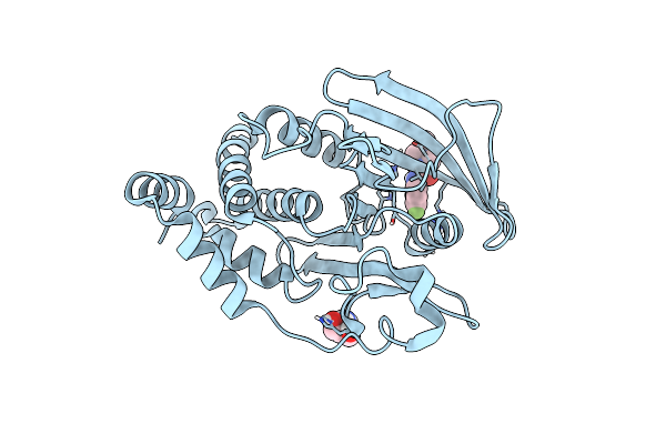
Crystal Structure Of Purine Nucleoside Phosphorylase Isoform 2 From Schistosoma Mansoni In Complex With 5-Fluoro-1,2-Dihydropyrimidin-2-One
Organism: Schistosoma mansoni
Method: X-RAY DIFFRACTION Resolution:1.92 Å Release Date: 2018-11-07 Classification: TRANSFERASE Ligands: DMS, DJP |
 |
Crystal Structure Of Purine Nucleoside Phosphorylase Isoform 2 From Schistosoma Mansoni In Complex With 2-Aminopyrimidin-5-Ol
Organism: Schistosoma mansoni
Method: X-RAY DIFFRACTION Resolution:2.00 Å Release Date: 2018-11-07 Classification: TRANSFERASE Ligands: DMS, DS1 |
 |
Crystal Structure Of Purine Nucleoside Phosphorylase Isoform 2 From Schistosoma Mansoni In Complex With (2R)-2-Amino-3-(Benzyloxy)Propan-1-Ol
Organism: Schistosoma mansoni
Method: X-RAY DIFFRACTION Resolution:1.42 Å Release Date: 2018-11-07 Classification: TRANSFERASE Ligands: DMS, SEM |
 |
Crystal Structure Of Purine Nucleoside Phosphorylase Isoform 2 From Schistosoma Mansoni In Complex With 1,2,5-Trimethyl-1H-Pyrrole-3-Carboxylic Acid
Organism: Schistosoma mansoni
Method: X-RAY DIFFRACTION Resolution:1.59 Å Release Date: 2018-11-07 Classification: TRANSFERASE Ligands: DMS, DS7 |
 |
Crystal Structure Of Purine Nucleoside Phosphorylase Isoform 2 From Schistosoma Mansoni In Complex With 1-(4-Amino-2-Hydroxyphenyl)Ethan-1-One
Organism: Schistosoma mansoni
Method: X-RAY DIFFRACTION Resolution:1.60 Å Release Date: 2018-11-07 Classification: TRANSFERASE Ligands: DMS, DSJ |
 |
Crystal Structure Of Purine Nucleoside Phosphorylase Isoform 2 From Schistosoma Mansoni In Complex With 2-{[(S)-Benzenesulfinyl]Methyl}Benzoic Acid
Organism: Schistosoma mansoni
Method: X-RAY DIFFRACTION Resolution:1.73 Å Release Date: 2018-11-07 Classification: TRANSFERASE Ligands: DMS, DTS |
 |
Crystal Structure Of Purine Nucleoside Phosphorylase Isoform 2 From Schistosoma Mansoni In Complex With 4-Chloro-6-Methylpyrimidin-2-Amine
Organism: Schistosoma mansoni
Method: X-RAY DIFFRACTION Resolution:1.56 Å Release Date: 2018-11-07 Classification: TRANSFERASE Ligands: DMS, DU7 |
 |
Crystal Structure Of Purine Nucleoside Phosphorylase Isoform 2 From Schistosoma Mansoni In Complex With 3-Methyl-1,2-Dihydropyridin-2-One
Organism: Schistosoma mansoni
Method: X-RAY DIFFRACTION Resolution:1.44 Å Release Date: 2018-10-17 Classification: TRANSFERASE/INHIBITOR Ligands: DMS, D4V |
 |
Pandda Analysis Group Deposition -- Crystal Structure Of Ptp1B In Complex With Compound_Fmopl000740A
Organism: Homo sapiens
Method: X-RAY DIFFRACTION Resolution:1.76 Å Release Date: 2018-10-10 Classification: HYDROLASE Ligands: AWD, TRS |