
Search Count: 37
 |
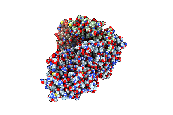
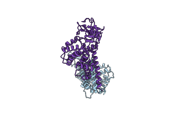
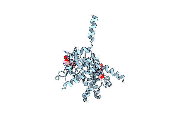
Crystal Structure Of Calcium/Calmodulin-Dependent Protein Kinase Type Ii Subunit Delta Complexed With Ribociclib
Organism: Homo sapiens
Method: X-RAY DIFFRACTION Resolution:2.35 Å Release Date: 2025-05-07 Classification: TRANSFERASE/TRANSFERASE INHIBITOR Ligands: 6ZZ |
 |
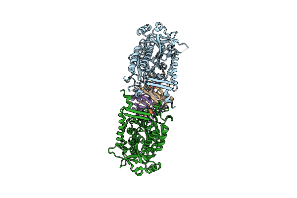
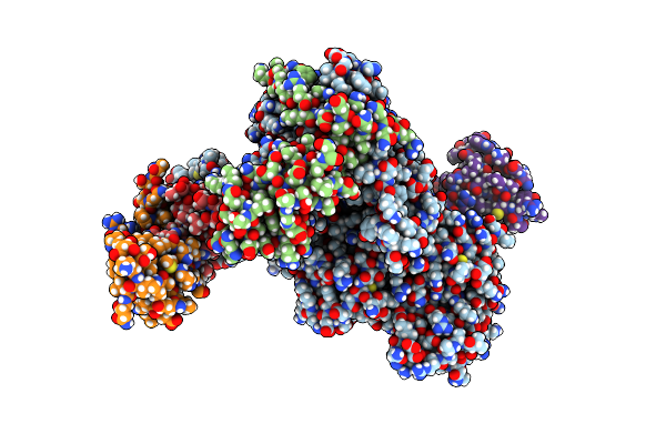
Crystal Structure Of Actin Capping Protein In Complex With A Fragment Of Legionella Pneumophila Ravb
Organism: Gallus gallus, Legionella pneumophila subsp. pneumophila (strain philadelphia 1 / atcc 33152 / dsm 7513)
Method: X-RAY DIFFRACTION Resolution:2.00 Å Release Date: 2025-05-07 Classification: PROTEIN BINDING Ligands: FMT |
 |
Organism: Severe acute respiratory syndrome coronavirus 2, Synthetic construct
Method: ELECTRON MICROSCOPY Release Date: 2025-03-12 Classification: VIRAL PROTEIN/RNA/INHIBITOR Ligands: ZN, A1AWQ, MN |
 |
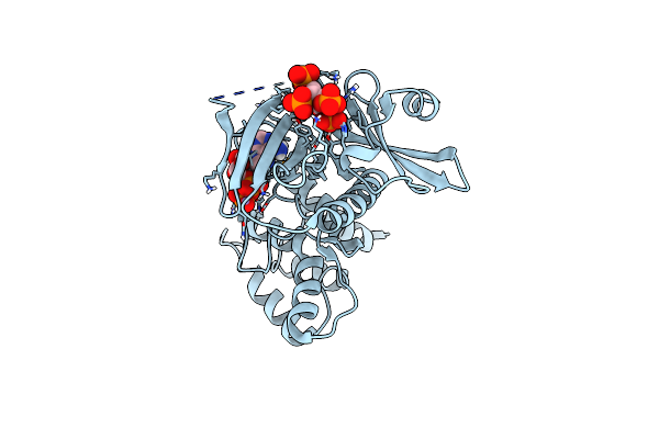
Organism: Escherichia coli
Method: ELECTRON MICROSCOPY Release Date: 2024-11-27 Classification: LIGASE/HYDROLASE Ligands: AMP |
 |
Organism: Escherichia coli
Method: ELECTRON MICROSCOPY Release Date: 2024-11-27 Classification: LIGASE Ligands: MN |
 |
Organism: Yasminevirus sp. gu-2018
Method: X-RAY DIFFRACTION Resolution:2.70 Å Release Date: 2024-10-02 Classification: LIGASE Ligands: AMP, NA, MG |
 |
Organism: Yasminevirus sp. gu-2018
Method: X-RAY DIFFRACTION Resolution:2.60 Å Release Date: 2024-10-02 Classification: LIGASE Ligands: ATP |
 |
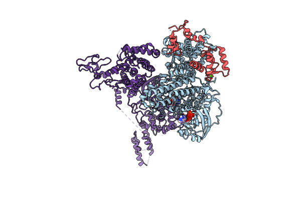
Organism: Homo sapiens
Method: ELECTRON MICROSCOPY Release Date: 2023-06-28 Classification: SIGNALING PROTEIN |
 |
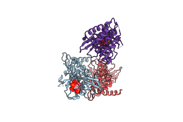
Organism: Homo sapiens
Method: ELECTRON MICROSCOPY Release Date: 2023-06-28 Classification: SIGNALING PROTEIN |
 |
Organism: Severe acute respiratory syndrome coronavirus 2
Method: ELECTRON MICROSCOPY Release Date: 2022-03-16 Classification: VIRAL PROTEIN Ligands: ZN, MN, POP |
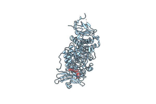 |
Structure Of The Starch Branching Enzyme I (Bei) Complexed With Maltododecaose From Oryza Sativa L
Organism: Oryza sativa subsp. japonica
Method: X-RAY DIFFRACTION Resolution:2.35 Å Release Date: 2021-11-17 Classification: TRANSFERASE/SUBSTRATE |
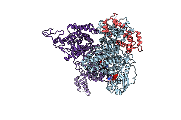 |
Organism: Legionella pneumophila, Homo sapiens
Method: ELECTRON MICROSCOPY Release Date: 2021-08-18 Classification: TRANSFERASE,HYDROLASE/LIGASE Ligands: AMP, MG, ATP, CA |
 |
Organism: Legionella pneumophila, Homo sapiens
Method: ELECTRON MICROSCOPY Release Date: 2021-08-18 Classification: TRANSFERASE/LIGASE Ligands: ATP, MG, NA, CA, AMP |
 |
Organism: Legionella pneumophila subsp. pneumophila
Method: X-RAY DIFFRACTION Resolution:3.20 Å Release Date: 2021-04-28 Classification: TRANSFERASE Ligands: ADP, MG |
 |
Organism: Legionella pneumophila
Method: X-RAY DIFFRACTION Resolution:2.10 Å Release Date: 2020-04-08 Classification: TRANSFERASE Ligands: EDO |
 |
Organism: Legionella pneumophila
Method: X-RAY DIFFRACTION Resolution:2.65 Å Release Date: 2020-04-08 Classification: TRANSFERASE Ligands: IHP |
 |
Organism: Legionella pneumophila
Method: X-RAY DIFFRACTION Resolution:1.85 Å Release Date: 2020-04-08 Classification: TRANSFERASE Ligands: IHP, ADP, MN |
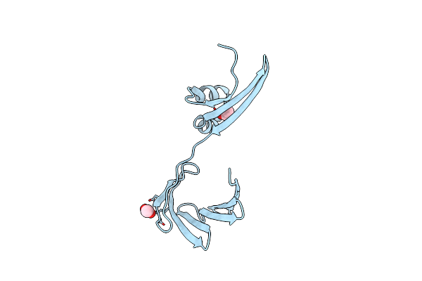 |
Crystal Structure Of The Apo Domain-Swapped Dimer Q108K:K40L:T51F Mutant Of Human Cellular Retinol Binding Protein Ii
Organism: Homo sapiens
Method: X-RAY DIFFRACTION Resolution:1.97 Å Release Date: 2019-10-16 Classification: CYTOSOLIC PROTEIN Ligands: ACT |
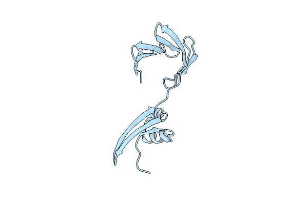 |
Crystal Structure Of The Apo Domain-Swapped Dimer Q108K:K40L:T51W Mutant Of Human Cellular Retinol Binding Protein Ii
Organism: Homo sapiens
Method: X-RAY DIFFRACTION Resolution:2.26 Å Release Date: 2019-10-16 Classification: LIPID BINDING PROTEIN Ligands: HOH |
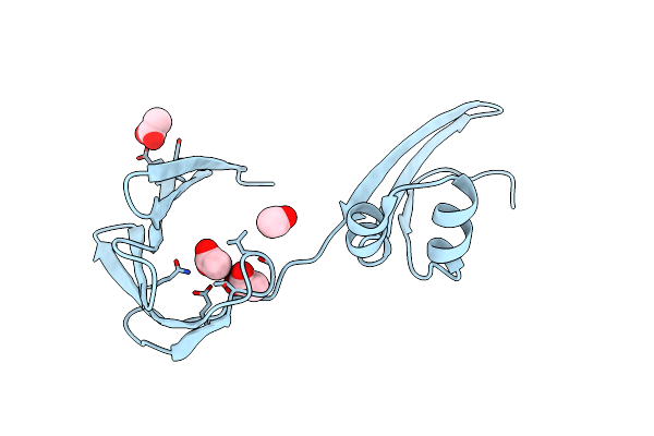 |
Crystal Structure Of The Apo Domain-Swapped Dimer Q108K:T51D Mutant Of Human Cellular Retinol Binding Protein Ii
Organism: Homo sapiens
Method: X-RAY DIFFRACTION Resolution:1.70 Å Release Date: 2019-10-16 Classification: LIPID BINDING PROTEIN Ligands: ACT |