
Search Count: 17
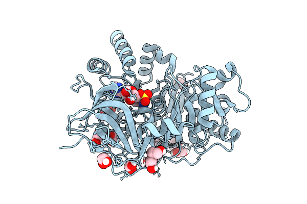 |
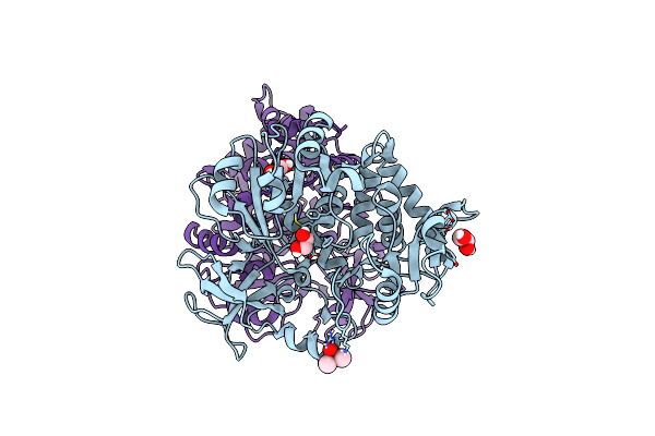
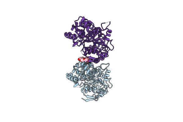
High-Resolution Crystal Structure Of Methyl-2,3-Diamino Propanoic Acid-Ams Inhibitor Bound Adenylation Domain (A3) From Sulfazecin Nonribosomal Peptide Synthetase Sulm
Organism: Paraburkholderia acidicola
Method: X-RAY DIFFRACTION Release Date: 2025-12-10 Classification: LIGASE Ligands: A1BVA, EDO, PEG |
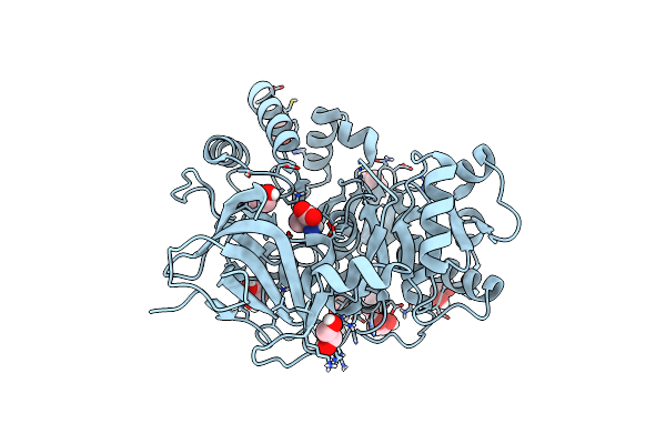 |
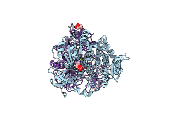
High-Resolution Crystal Structure Of 2,3-Diamino Propanoic Acid Bound Adenylation Domain (A3) From Sulfazecin Nonribosomal Peptide Synthetase Sulm
Organism: Paraburkholderia acidicola
Method: X-RAY DIFFRACTION Resolution:1.55 Å Release Date: 2025-05-28 Classification: LIGASE Ligands: EDO, DPP |
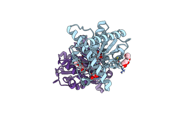 |
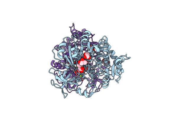
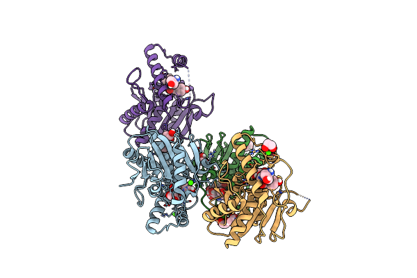
Thioesterase Domain Structure From Sulfazecin Biosynthetic Nonribosomal Peptide Synthetase Sulm
Organism: Paraburkholderia acidicola
Method: X-RAY DIFFRACTION Resolution:1.90 Å Release Date: 2024-08-07 Classification: HYDROLASE Ligands: EDO, NA |
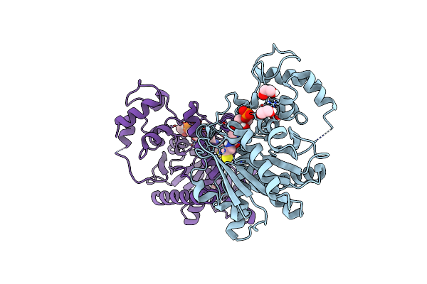 |
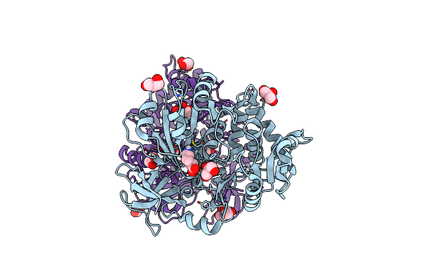
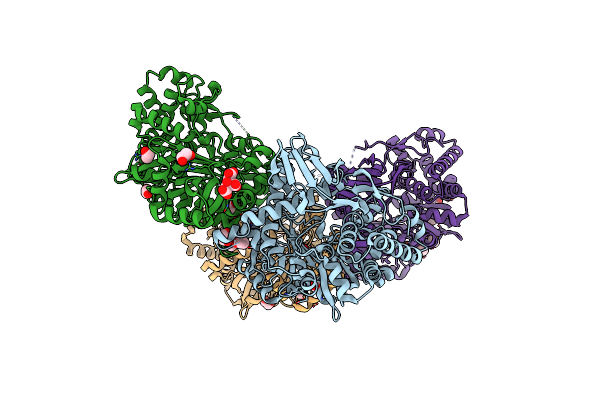
Holo-Pcp-Thioesterase Di-Domain Structure From The Sulfazecin Biosynthetic Nonribosomal Peptide Synthetase, Sulm
Organism: Paraburkholderia acidicola
Method: X-RAY DIFFRACTION Resolution:2.70 Å Release Date: 2024-08-07 Classification: HYDROLASE Ligands: PNS, EDO |
 |
Adenylation Domain Structure From Nrps-Like Delta-Poly-L-Ornithine Synthetase
Organism: Acinetobacter baumannii ab307-0294
Method: X-RAY DIFFRACTION Resolution:2.20 Å Release Date: 2023-10-04 Classification: LIGASE Ligands: EDO, CAC |
 |
Adenylation Domain Structure From Nrps-Like Delta-Poly-L-Ornithine Synthetase (L-Ornithine Bound)
Organism: Acinetobacter baumannii ab307-0294
Method: X-RAY DIFFRACTION Resolution:2.30 Å Release Date: 2023-10-04 Classification: LIGASE Ligands: ORN, GOL |
 |
Adenylation Domain Structure From Nrps-Like Delta-Poly-L-Ornithine Synthetase (D-Ornithine Bound)
Organism: Acinetobacter baumannii ab307-0294
Method: X-RAY DIFFRACTION Resolution:2.51 Å Release Date: 2023-10-04 Classification: LIGASE Ligands: GOL, MG, ORD |
 |
Adenylation Domain Structure From Nrps-Like Delta-Poly-L-Ornithine Synthetase (L-Lysine Bound)
Organism: Acinetobacter baumannii ab307-0294
Method: X-RAY DIFFRACTION Resolution:2.49 Å Release Date: 2023-10-04 Classification: LIGASE Ligands: GOL, LYS |
 |
Crystal Structure Of Desd, The Desferrioxamine Synthetase From The Streptomyces Griseoflavus Ferrimycin Biosynthetic Pathway
Organism: Streptomyces griseoflavus
Method: X-RAY DIFFRACTION Resolution:2.85 Å Release Date: 2022-07-06 Classification: LIGASE Ligands: GOL, SO4 |
 |
Crystal Structure Of Atp Bound Desd, The Desferrioxamine Synthetase From The Streptomyces Griseoflavus Ferrimycin Biosynthetic Pathway
Organism: Streptomyces griseoflavus
Method: X-RAY DIFFRACTION Resolution:2.30 Å Release Date: 2022-07-06 Classification: LIGASE Ligands: SO4, ATP, MG, B3P, MPD |
 |
Crystal Structure Of Amp+Ppi Bound Desd, The Desferrioxamine Synthetase From The Streptomyces Griseoflavus Ferrimycin Biosynthetic Pathway
Organism: Streptomyces griseoflavus
Method: X-RAY DIFFRACTION Resolution:2.89 Å Release Date: 2022-07-06 Classification: LIGASE Ligands: GOL, PPV, AMP, SO4, MG |
 |
Crystal Structure Of Hsc-Ams Bound Desd, The Desferrioxamine Synthetase From The Streptomyces Griseoflavus Ferrimycin Biosynthetic Pathway
Organism: Streptomyces griseoflavus
Method: X-RAY DIFFRACTION Resolution:2.50 Å Release Date: 2022-07-06 Classification: LIGASE/INHIBITOR Ligands: GOL, I3U, LMS, PO4, MG, SO4 |
 |
Crystal Structure Of Desd, The Desferrioxamine Synthetase From The Streptomyces Violaceus Salmycin Biosynthetic Pathway
Organism: Streptomyces violaceus
Method: X-RAY DIFFRACTION Resolution:2.30 Å Release Date: 2022-07-06 Classification: LIGASE Ligands: EDO, MG, FLC |
 |
Crystal Structure Of Photinus Pyralis Luciferase Pps6 Mutant In Complex With Dlsa
Organism: Photinus pyralis
Method: X-RAY DIFFRACTION Resolution:2.75 Å Release Date: 2019-10-16 Classification: LIGASE Ligands: SO4, DYD, EDO, SLU |
 |
A High-Resolution Crystal Structure Of Nocb Thioesterase Domain From Nocardicin Cluster
Organism: Nocardia uniformis subsp. tsuyamanensis
Method: X-RAY DIFFRACTION Resolution:1.94 Å Release Date: 2019-09-11 Classification: HYDROLASE Ligands: SO4, GOL |
 |
A High-Resolution Crystal Structure Of Covalent Complex Of Nocb Thioesterase Domain With Fluorophosphonate Nocardicin G Analog
Organism: Nocardia uniformis subsp. tsuyamanensis
Method: X-RAY DIFFRACTION Resolution:1.99 Å Release Date: 2019-09-11 Classification: HYDROLASE Ligands: GOL, MUY, CA |
 |
High Resolution Crystal Structure Of Reductase (R) Domain Of Nonribosomal Peptide Synthetase From Mycobacterium Tuberculosis
Organism: Mycobacterium tuberculosis
Method: X-RAY DIFFRACTION Resolution:1.81 Å Release Date: 2016-01-13 Classification: LIGASE Ligands: XPE |