
Search Count: 7
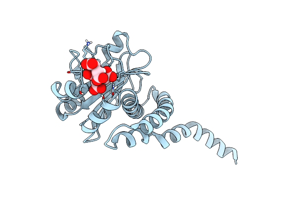 |
Structure Of Bacterial Sylf Domain Containing Protein, Beta Cell Expansion Factor A (Befa)
Organism: Aeromonas veronii
Method: X-RAY DIFFRACTION Resolution:1.27 Å Release Date: 2022-07-20 Classification: SIGNALING PROTEIN Ligands: CIT |
 |
Periplasmic Portion Of The Helicobacter Pylori Chemoreceptor Tlpb With Urea Bound
Organism: Helicobacter pylori
Method: X-RAY DIFFRACTION Resolution:1.38 Å Release Date: 2012-06-27 Classification: MEMBRANE PROTEIN Ligands: URE, SO4, PEG, GOL |
 |
Periplasmic Portion Of The Helicobacter Pylori Chemoreceptor Tlpb With Acetamide Bound
Organism: Helicobacter pylori
Method: X-RAY DIFFRACTION Resolution:1.40 Å Release Date: 2012-06-27 Classification: MEMBRANE PROTEIN Ligands: ACM, SO4, GOL |
 |
Periplasmic Portion Of The Helicobacter Pylori Chemoreceptor Tlpb With Formamide Bound
Organism: Helicobacter pylori
Method: X-RAY DIFFRACTION Resolution:1.42 Å Release Date: 2012-06-27 Classification: MEMBRANE PROTEIN Ligands: ARF, SO4, GOL |
 |
Periplasmic Portion Of The Helicobacter Pylori Chemoreceptor Tlpb With Hydroxyurea Bound
Organism: Helicobacter pylori
Method: X-RAY DIFFRACTION Resolution:1.42 Å Release Date: 2012-06-27 Classification: MEMBRANE PROTEIN Ligands: NHY, SO4, GOL |
 |
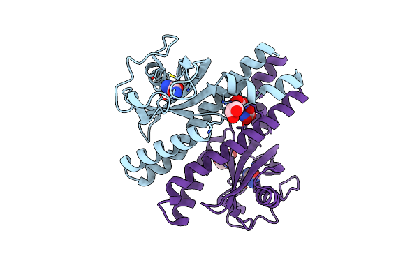
Revised, Rerefined Crystal Structure Of Pdb Entry 2Qhk, Methyl Accepting Chemotaxis Protein
Organism: Vibrio parahaemolyticus
Method: X-RAY DIFFRACTION Resolution:1.90 Å Release Date: 2012-05-30 Classification: SIGNALING PROTEIN Ligands: PYR |
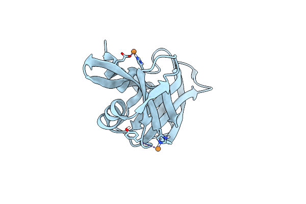 |
Organism: Bos taurus
Method: X-RAY DIFFRACTION Resolution:2.00 Å Release Date: 1998-05-27 Classification: HYDROLASE (PHOSPHORIC DIESTER) Ligands: CU |