
Search Count: 6
 |
Organism: Escherichia coli (strain k12)
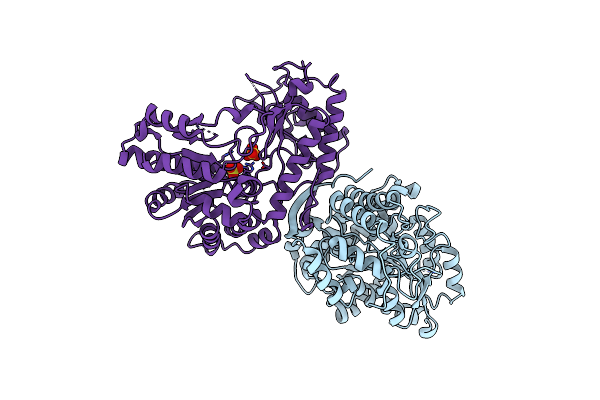
Method: X-RAY DIFFRACTION Resolution:1.89 Å Release Date: 2015-12-09 Classification: LYASE Ligands: CL, MG, EPE, NA, CA |
 |
Organism: Escherichia coli
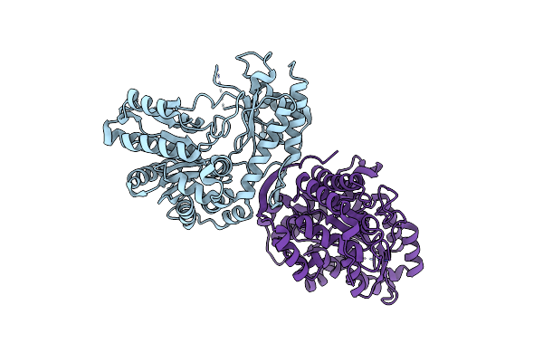
Method: X-RAY DIFFRACTION Resolution:3.20 Å Release Date: 2014-12-17 Classification: LYASE Ligands: SO4 |
 |
Organism: Escherichia coli
Method: X-RAY DIFFRACTION Resolution:2.89 Å Release Date: 2014-12-17 Classification: LYASE |
 |
Organism: Escherichia coli
Method: X-RAY DIFFRACTION Resolution:2.00 Å Release Date: 2008-06-10 Classification: LYASE Ligands: CL, MG |
 |
Organism: Escherichia coli
Method: X-RAY DIFFRACTION Resolution:1.90 Å Release Date: 2008-06-10 Classification: LYASE Ligands: CL, MG |
 |
Crystal Structure Of The Apo Form Of E. Coli Tryptophanase At 1.9 A Resolution
Organism: Escherichia coli
Method: X-RAY DIFFRACTION Resolution:1.90 Å Release Date: 2007-02-20 Classification: LYASE Ligands: CL, MG, EPE |