
Search Count: 29
 |
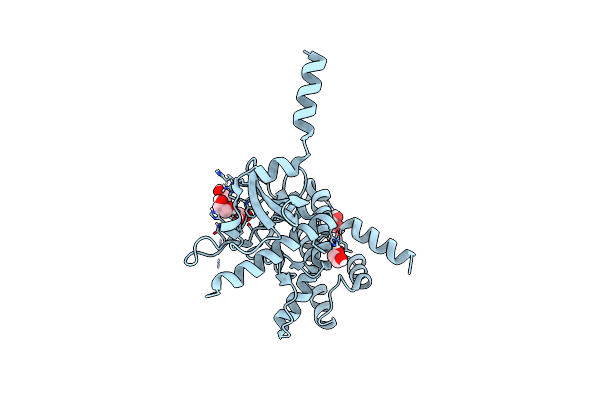
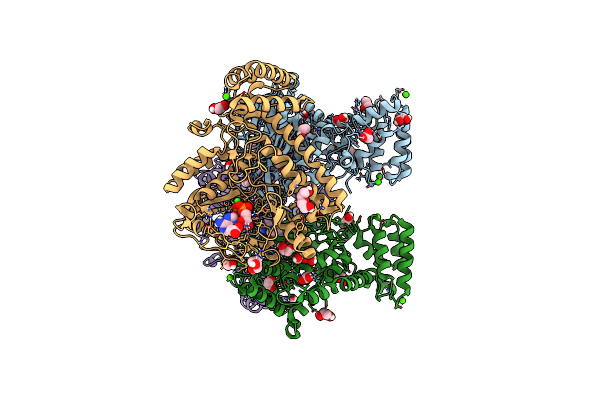
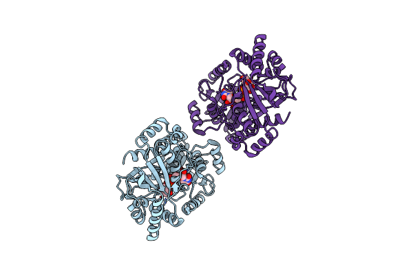
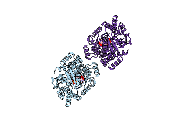
Crystal Structure Of Calcium/Calmodulin-Dependent Protein Kinase Type Ii Subunit Delta Complexed With Ribociclib
Organism: Homo sapiens
Method: X-RAY DIFFRACTION Resolution:2.35 Å Release Date: 2025-05-07 Classification: TRANSFERASE/TRANSFERASE INHIBITOR Ligands: 6ZZ |
 |
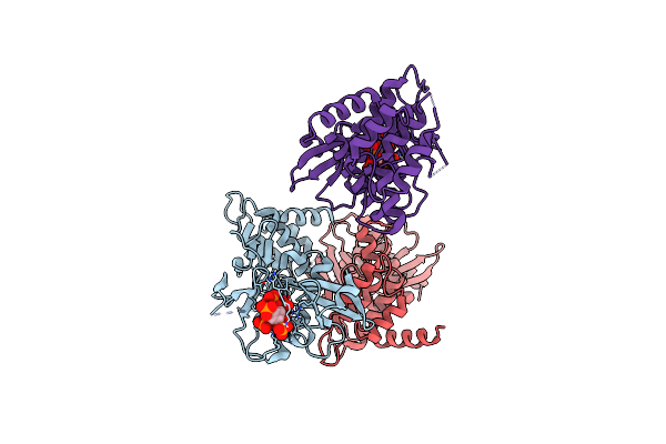
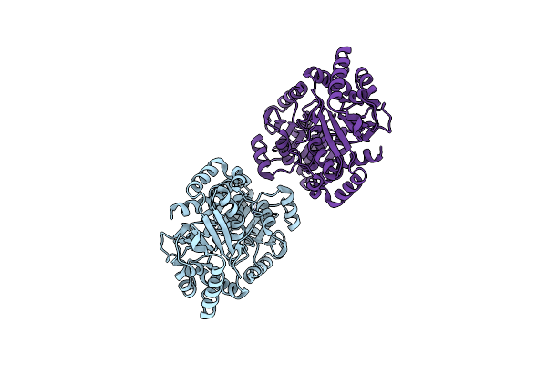
Organism: Legionella pneumophila
Method: X-RAY DIFFRACTION Resolution:2.10 Å Release Date: 2020-04-08 Classification: TRANSFERASE Ligands: EDO |
 |
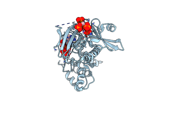
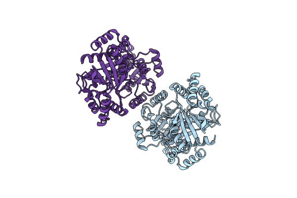
Organism: Legionella pneumophila
Method: X-RAY DIFFRACTION Resolution:2.65 Å Release Date: 2020-04-08 Classification: TRANSFERASE Ligands: IHP |
 |
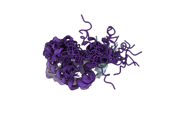
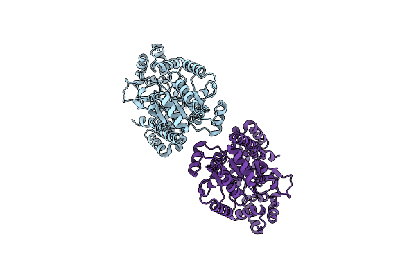
Organism: Legionella pneumophila
Method: X-RAY DIFFRACTION Resolution:1.85 Å Release Date: 2020-04-08 Classification: TRANSFERASE Ligands: IHP, ADP, MN |
 |
Organism: Ewingella americana
Method: X-RAY DIFFRACTION Resolution:2.47 Å Release Date: 2019-09-11 Classification: TRANSFERASE Ligands: EDO, SO4 |
 |
Organism: Ewingella americana
Method: X-RAY DIFFRACTION Resolution:1.89 Å Release Date: 2019-09-11 Classification: TRANSFERASE Ligands: ANP, EDO |
 |
 |
Organism: Pseudomonas syringae
Method: X-RAY DIFFRACTION Resolution:2.27 Å Release Date: 2018-10-03 Classification: TRANSFERASE Ligands: MG, CA, CL, ANP, EDO, PEG, ACT |
 |
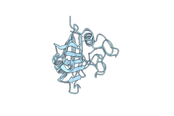 |
Organism: Triticum aestivum
Method: X-RAY DIFFRACTION Resolution:1.25 Å Release Date: 2013-03-27 Classification: ISOMERASE |
 |
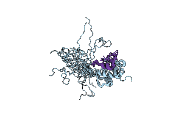
Structural And Biochemical Characterization Of A Cytosolic Wheat Cyclophilin Tacypa-1
Organism: Triticum aestivum, Tolypocladium inflatum
Method: X-RAY DIFFRACTION Resolution:1.20 Å Release Date: 2013-03-27 Classification: ISOMERASE/INHIBITOR |
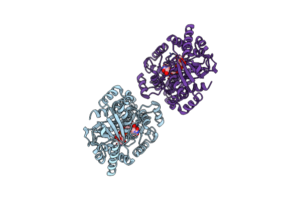 |
Crystal Structure Of Trehalose Synthase Tret Mutant E326A From P. Horikoshii In Complex With Udpg
Organism: Pyrococcus horikoshii
Method: X-RAY DIFFRACTION Resolution:2.50 Å Release Date: 2011-03-16 Classification: BIOSYNTHETIC PROTEIN Ligands: UPG |
 |
Crystal Structure Of Trehalose Synthase Tret Mutant E326A From P. Horikoshii In Complex With Udpg
Organism: Pyrococcus horikoshii
Method: X-RAY DIFFRACTION Resolution:2.50 Å Release Date: 2011-03-16 Classification: BIOSYNTHETIC PROTEIN Ligands: UPG |
 |
Organism: Escherichia coli
Method: X-RAY DIFFRACTION Resolution:2.90 Å Release Date: 2011-01-26 Classification: CHAPERONE |
 |
Organism: Homo sapiens
Method: X-RAY DIFFRACTION Resolution:2.70 Å Release Date: 2010-12-29 Classification: HYDROLASE |
 |
Glycoside Hydrolase Family 77 4-Alpha-Glucanotransferase From Thermus Brockianus
Organism: Thermus brockianus
Method: X-RAY DIFFRACTION Resolution:2.36 Å Release Date: 2010-10-27 Classification: TRANSFERASE Ligands: PO4, SO4, INS |
 |
Organism: Pyrococcus horikoshii
Method: X-RAY DIFFRACTION Resolution:2.20 Å Release Date: 2010-10-13 Classification: BIOSYNTHETIC PROTEIN |
 |
Crystal Structure Of Trehalose Synthase Tret From P.Horikoshi Produced By Soaking In Trehalose
Organism: Pyrococcus horikoshii
Method: X-RAY DIFFRACTION Resolution:2.20 Å Release Date: 2010-10-13 Classification: ISOMERASE |
 |
Crystal Structure Of Trehalose Synthase Tret From P.Horikoshii (Seleno Derivative)
Organism: Pyrococcus horikoshii
Method: X-RAY DIFFRACTION Resolution:2.47 Å Release Date: 2010-10-13 Classification: BIOSYNTHETIC PROTEIN |
 |
Crystal Structure Of Trehalose Synthase Tret Mutant E326A From P. Horishiki In Complex With Udp
Organism: Pyrococcus horikoshii
Method: X-RAY DIFFRACTION Resolution:2.50 Å Release Date: 2010-10-13 Classification: SUGAR BINDING PROTEIN Ligands: UDP |