
Search Count: 22
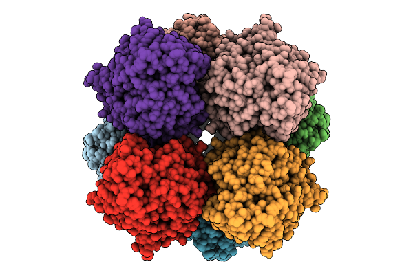 |
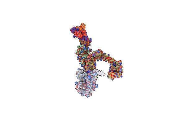
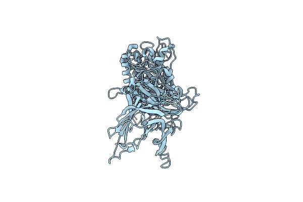
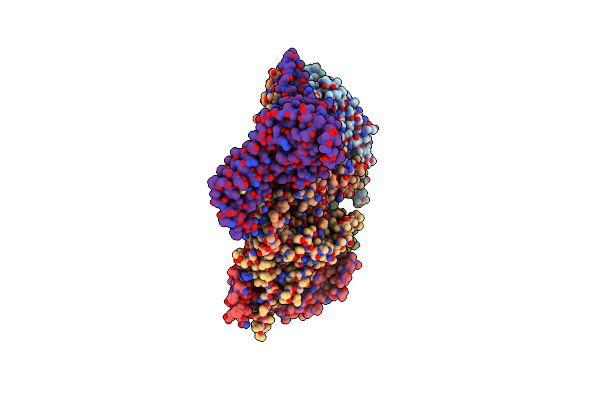
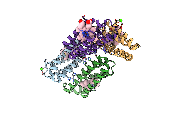
Structure Of Lara-Like Nickel-Pincer Nucleotide Cofactor-Utilizing Enzyme With A Single Catalytic Histidine Residue From Streptococcus Plurextorum
Organism: Streptococcus plurextorum
Method: ELECTRON MICROSCOPY Release Date: 2025-09-24 Classification: ISOMERASE Ligands: NI, 4EY |
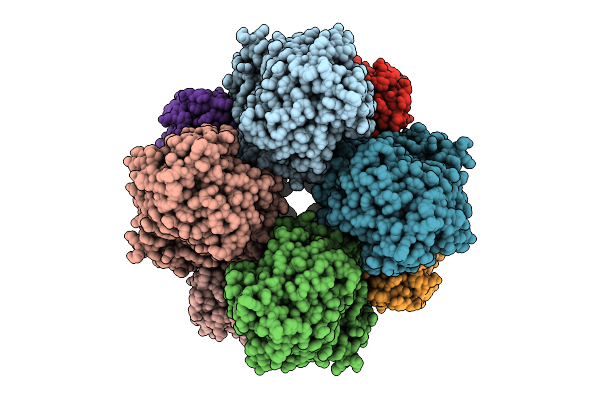 |
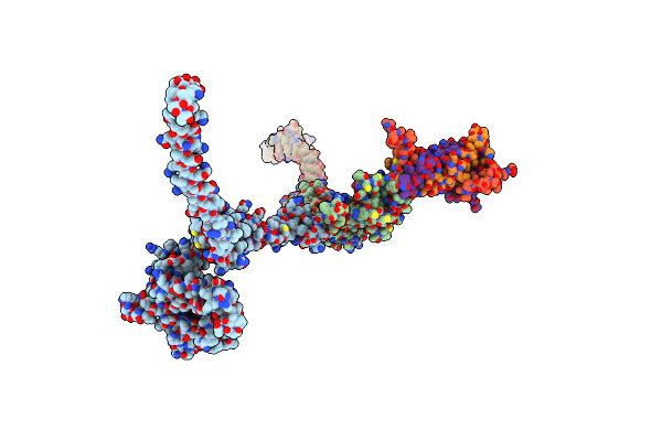
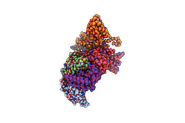
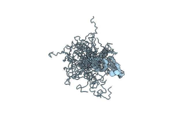
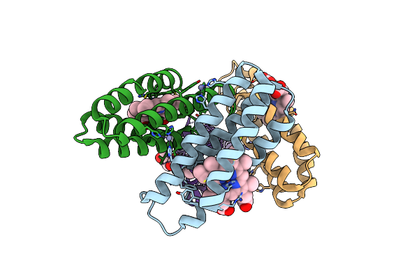
Structures Of Lara-Like Nickel-Pincer Nucleotide Cofactor-Utilizing Enzyme With A Single Catalytic Histidine Residue From Blautia Wexlerae
Organism: Blautia wexlerae
Method: ELECTRON MICROSCOPY Release Date: 2025-09-17 Classification: ISOMERASE |
 |
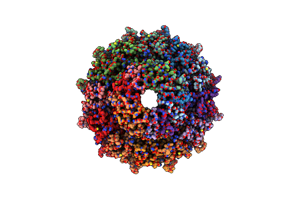
Organism: Escherichia phage ms2
Method: ELECTRON MICROSCOPY Release Date: 2025-08-06 Classification: VIRUS LIKE PARTICLE |
 |
Organism: Escherichia phage ms2
Method: ELECTRON MICROSCOPY Release Date: 2025-08-06 Classification: VIRUS LIKE PARTICLE |
 |
Organism: Shigella virus moo19
Method: ELECTRON MICROSCOPY Release Date: 2025-01-15 Classification: VIRUS |
 |
Organism: Shigella virus moo19
Method: ELECTRON MICROSCOPY Release Date: 2025-01-15 Classification: VIRUS |
 |
Organism: Shigella virus moo19
Method: ELECTRON MICROSCOPY Release Date: 2025-01-15 Classification: VIRAL PROTEIN |
 |
Organism: Shigella phage b2
Method: ELECTRON MICROSCOPY Release Date: 2025-01-15 Classification: VIRUS |
 |
Organism: Shigella phage b2
Method: ELECTRON MICROSCOPY Release Date: 2025-01-15 Classification: VIRUS |
 |
Organism: Shigella phage b2
Method: ELECTRON MICROSCOPY Release Date: 2025-01-15 Classification: VIRAL PROTEIN |
 |
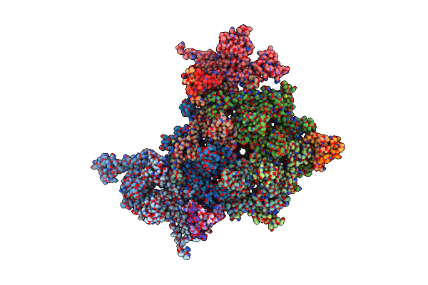
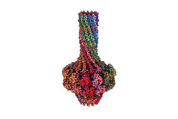
A Vibrio Cholerae Viral Satellite Enables Efficient Horizontal Transfer By Using An External Scaffold To Assemble Hijacked Coat Proteins Into Small Capsids
Organism: Vibrio phage icp1_2011_a, Vibrio cholerae
Method: ELECTRON MICROSCOPY Release Date: 2024-01-17 Classification: VIRUS LIKE PARTICLE |
 |
Organism: Shigella phage buco
Method: ELECTRON MICROSCOPY Release Date: 2023-10-18 Classification: VIRUS |
 |
Organism: Shigella phage buco
Method: ELECTRON MICROSCOPY Release Date: 2023-09-27 Classification: VIRUS |
 |
Organism: Shigella phage buco
Method: ELECTRON MICROSCOPY Release Date: 2023-09-27 Classification: VIRUS |
 |
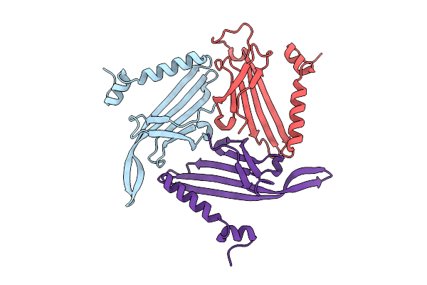
Nmr Solution Structure Of The Monomeric Form Of The Phage L Decoration Protein
Organism: Enterobacteria phage l
Method: SOLUTION NMR Release Date: 2019-04-17 Classification: VIRAL PROTEIN |
 |
Organism: Enterobacteria phage p22
Method: X-RAY DIFFRACTION Resolution:3.30 Å Release Date: 2017-02-08 Classification: VIRAL PROTEIN |
 |
Organism: Enterobacteria phage p22
Method: X-RAY DIFFRACTION Resolution:7.00 Å Release Date: 2017-02-08 Classification: VIRAL PROTEIN |
 |
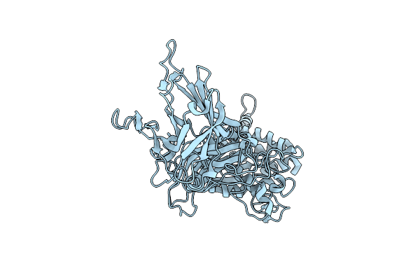
Crystal Structure Of An Engineered Cytochrome Cb562 That Forms 1D, Zn-Mediated Coordination Polymers
Organism: Escherichia coli
Method: X-RAY DIFFRACTION Resolution:2.00 Å Release Date: 2012-07-04 Classification: METAL BINDING PROTEIN Ligands: HEM, ZN, CA |
 |
Crystal Structure Of An Engineered Cytochrome Cb562 That Forms 2D, Zn-Mediated Sheets
Organism: Escherichia coli
Method: X-RAY DIFFRACTION Resolution:2.30 Å Release Date: 2012-07-04 Classification: METAL BINDING PROTEIN Ligands: HEM, ZN |
 |
Structural Characterization Of The Dual Glycan Binding Adeno-Associated Virus Serotype 6
Organism: Adeno-associated virus - 6
Method: X-RAY DIFFRACTION Resolution:3.00 Å Release Date: 2011-03-23 Classification: VIRUS Ligands: CYT, OAH |